Introduction
Spinal tumor is an abnormal mass of tissue within or surrounding spinal cord and spinal column. Intraspinal tumours can originate from the spinal cord, filum terminale, nerve roots, meninges, intraspinal vessels, sympathetic chain and vertebrae. These tumors can be benign or malignant, primary or secondary, and can result in serious morbidity. Role of squash cytology for rapid intraoperative cytological diagnosis of space‑ occupying lesions (SOLs) in the brain is well established and has been an important diagnostic tool over the last few decades.
However, its role in spinal lesions is unexplored. Recently, the rapid cytological diagnosis for spinal lesions is also gaining importance, which is diagnostically challenging for neuropathologist. Intraoperative diagnosis helps surgeons to decide on optimal line of management for various spinal lesions without having to wait for routine paraffin‑ embedded sections. The aim of this study was to compare the efficacy of intraoperative squash smear cytology with histopathological diagnosis .
Materials and Methods
This study was conducted over a period of 15 months in the Department of Pathology collaborating with Department of Neurosurgery in a tertiary care hospital. A total of 20 cases of spinal tumors were included in this study. The samples were sent immediately to pathology laboratory in isotonic saline for processing. Gross examination was done to evaluate nature of spread. A small proportion of the biopsy sample was crushed between two slides and smear prepared. The smears were immediately put in 95% ethyl alcohol for 2 minutes for wet fixation and stained with rapid H and E stain and air dried smears with toluedine blue. Remaining tissue processed for paraffin sections for histopathological report and immunohistochemistry was done wherever necessary. The radiological findings and clinical history in detail was available during the intraoperative diagnosis. Rest of the sample was immediately fixed for staining with Hematoxylin and Eosin (H and E). Smears were mounted with DPX. Final diagnosis was done on histopathological and compared with Initial cytological diagnosis .
Results
A total of 20 cases of spinal space occupying lesions were studied by squash cytology and histopathology. The mean age of presentation was 40.3 years. We had 13 male patients and 7 female patients. All 4 cases of metastatic follicular carcinoma of thyroid were, however, seen in female patients. Squash cytological diagnosis was compared with final histological diagnosis in all the cases, which was considered to be the gold standard. The spectrum of spinal lesions in our series has been summarized in Table 1.
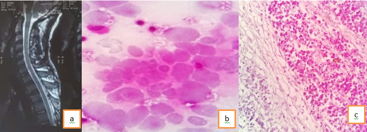
The commonest lesion in our series was Metastatic Adenocarcinoma [Figure 1 a-c]
(30 % of the total cases) followed by Schwannoma and Metastasis foll icular carcinoma of thyroid (20%) [Figure 2 a-c]. The other tumors were Neuroblastoma, ependymoma, PNET metastatic adenosquamous carcinoma and metastasis from seminoma. The cytological diagnosis was made and categorized as benign or malignant provisionally and final histolopathological diagnosis was made according to WHO classification and correlated. [Table 2 a]
Discordant cases is shown in Table 2 b. There was 1 discordant case. The final histopathological diagnosis was also correlated with radiologi cal diagnosis, shown in Table 3
A case of spinal metastasis was misdiagnosed as plasmacytoma radiologically, diagnosed as Metastasis Adenocarcinoma and advised to investigate for possibility of thyroid malignancy on squash cytology and later confirmed as Metastasic deposits from Follicular carcinoma of thyroid. (Figure 1) One case was diagnosed as potts spine radiologically, on squash cytology only fibroadipose tissue and inflammation was seen , it was diagnosed as Metastatic Adenocarcinoma from Prostate. [Figure 2 a-c] One case was diagnosed as Meningioma / Aneurysmal bone cyst radiologically, was diagnosed as Metastatic lesion on squash cytology and histopathology. Four cases were given differential diagnosis of meningioma / neurofibroma, diagnosed as Schwannoma on squash cytology and confirmed as Schwannoma on histopathology. A complete cytohistological correlation was seen in cases of Ependymoma [Figure 3 a-c], Neuroblastoma, PNET, Metastasis from operated case for testicular tumor [Figure 4 a-c] and 3 cases of Metastasis from Follicular carcinoma Thyroid .
Immunohistochemistry of Cytokeratin, EMA, GFAP and ki 67 was done wherever necessary for confirmation of diagnosis.
In our study, diagnosis by squash preparation was accurate in 95 % of cases.
Diagnostic accuracy by squash cytology was accurate for 11 out of 12 cases of metastatic lesions.
Figure 1
a- CT :-Plasmacytoma / Metastatic; b Squash diagnosis: low power- Metastatic adenocarcinoma; c- H/P high power shows cells arranged in microfollicular and macrofollicular pattern containing colloid - Metastatic Follicular Thyroid Carcinoma.

Figure 2
a Central compression fracture of the body of D3 vertebrae. Lesion is se en causing indentation on spinal cord from anterolateral aspect resulting in compression of the cord. Considering above features possibility of tuberculosis merits prime consideration however possibility of metastasis cannot be ruled out ; 2b – Squash cytology on high power – shows inflammatory cells and fibroconnective tissue – Inflammatory lesion ; 2c – H/p - High power shows fibrocollagenous tissue , inflammation and foci of metastatic adenocarcinoma

Figure 3
a – Well defined intra medullary contrast enhancing lesions measuring1.6x1.3x.3cm at L1and L2 vertebral boby displacing nerve root, predomnantly involving conus medullaris / filum terminale. Consistent with ependymoma. D.D includes astrocytoma .3b – Squash diagnosis- 10x Tumor cells arranged in papillary pattern , arround the bl ood vessels, having myxoid core 3c- H/P on high power – cuboidal to elongated tumour cells radially arranged in a papillary manner around vascularized stromal cores and also seen are Myxoid perivascular pseudorosettes.

Figure 4
a- Focal lesion in L1 – L4 vertebra bodies with anterior epidural soft tissue component causing compressionin nerve root at L1- L4 vertebra - Most likely Metastatic lesion Fig 4b- Squash cytology – high power showing large pleomorphic cells with lymphocytes. Fig 4c – H/p – low power showing nest of cells separated by fibrous septa infiltrate with mononuclear cell mostly lymphocytes. Tumor cells pleomorphic hyper chromatic nuclei with eosinophilic cytoplasm. Some cell are showing perinuclear clearing of cytoplasm.

Table 1
Histopathological spectrum of spinal tumors
Table 2
Correlation of Histopathological Versus cytological Diagnosis is shown in Table 2a
Table 3
Radiological and histopathological correlation
Discussion
The intraoperative diagnosis by cytology was first started in 1930 by Eisenhardt and Cushing, followed by Badt in 1937.1,2 With the introduction of computed tomography and magnetic
resonance imaging‑guided stereotactic biopsies, the importance of squash cytology is increasing day‑by‑day.3 The main purpose of intraoperative squash cytology is to provide surgeons adequate details about the lesion, so that the extent of surgery can be optimized and further therapeutic approach can be modified in an individualized manner.4,5 It is utmost important to classify tumors as prognosis is partly dependant on cell type. Diagnosing brain lesions by squash cytology has been well documented but less studies are available about its utility in spinal tumors. Therefore, the aim of our study was mainly to assess role of squash cytology in spinal tumors and correlate with histopathology.
The present study observed that spinal tumors were distributed in all ranges similar to studies done by Karn Bhardwaj et al, Mousumi kar et al.6,7 Peak incidence of tumors was in the middle life.
Majority of the cases in our study were of Metastatic lesions, The spinal column is the most common site for bone metastasis. The spine is the second-most-common location for metastatic disease to the CNS in patients with malignancies, after brain.
Estimates indicate that at least 30 percent and as high as 70 percent of patients with cancer will experience spread of cancer to their spine. Common primary cancers that spread to the spine are lung, breast and prostate Prostate cancer has 3 notable features, it does not enter into brain, it had predilection for dura and it can cytologically be relatively bland, all these features are shared with meningioma.8 Metastasis produce mass effect by their size and by the edema they generate , unchecked this effect can rapidly lead to herniation and death. Diagnostic accuracy was 91.6 % in Metastatic lesions in our cases similar to studies done earlier.9,10,11,12
Schwannomas are major extra-axial intradural tumor .These tumors produce a dense collagenous matrix and envelope individual cells in a tenatious reticulin network. Cyto histopathological correlation was 100 % in case of schwannomas, In a study done by Mousumi kar et al , majority of the tumors were of Schwannomas but in our study schwannomas were the second common tumors.7
Ependymomas of spinal cord is most common intramedullary spinal tumor of adults.
In our study diagnostic accuracy was 100 % , similar to study done by Mousumi kar et al.7
In the study by Mousumi kar et al, the diagnostic sensitivity of squash preparation was 95.75%. They further documented diagnostic accuracy of squash cytology in individual tumors. Diagnostic accuracy for schwa nnoma was 92.3% which was in approximation with our study.
In a similar study, squash cytology done on central nervous system lesions (both on spinal and brain SOLs) documented 88% overall diagnostic accuracy.3
In a study done on both neoplastic and nonneoplastic brain lesions, diagnostic accuracy of squash cytology was found to be 93.3%.13
Although diagnostic accuracy rates are higher for brain SOLs than spinal lesions in various studies, we found that the results of our series based on spinal SOLs alone are also comparable. There is very less data available on spinal lesions alone.
Squash cytology provides invaluable cytological information such as cytoplasmic and nuclear details along with important cellular architecture such as whorls rosettes, etc.14,15,16
Artifacts such as crushing, overstretching, and cellular overlapping can impose problems in making an accurate diagnosis by squash.
Tanycytic ependymoma is a common tumor of the spine which has relatively few perivascular rosettes, and the presence of spindle ‑shaped cells can be misleading. It can be mistaken for
pilocytic astrocytoma or schwannoma. In our series, it was diagnosed correctly.
A completre 100% cytohistological correlation was seen in Neuroblastoma and PNET similar to study done by Kishore Sanjeev et al.
Almost all studies reviewed for present work have reported high accuracy rate of intraoperative squash smear diagnosis of intracranial and spinal cord tumors as approximately to 90% and higher9,10,11,12
Conclusion
In conclusion this study shows a high degree of cytohistological correlation . Squash cytology is an important intraoperative diagnostic tool in management of brain tumors. Its role in spinal lesion is not fully established. In our study we found that the diagnostic accuracy of spinal lesions was comparable with brain SOLs and it was found to be high.
Also important in case of spinal tumors to bear in mind the radiological finding, age distribution, and anatomical location to come to an accurate cytological diagnosis.
