Introduction
Erlich is credited with the first liver aspiration in 1883 and subsequently first percutaneous liver biopsy for diagnostic purposes was reported in 1923. The technique has been modified since then and proved to be a revolution in the field of hepatology.1,2 It is cornerstone in the evaluation and management of patients with liver disease and has long been considered to be an integral component of the clinician’s diagnostic armamentarium.3
The value of liver biopsy is not merely to determine the degree of fibrosis, rather it draws a detailed map for many important histological findings such as the degree of inflammation, nature of inflammatory cells, distribution of inflammation, status of bile ducts, vasculature, presence of steatosis and deposition and infiltration of liver with different materials like iron, copper, etc. Undoubtedly, this otherwise unobtainable information regarding the structural integrity of liver parenchyma, degree and type of injury and the host response, has a clear impact on the diagnosis, prognosis and response to treatment.2
The ease and low mortality and relatively low morbidity of this procedure has made it to be widely used.1 Thus, liver biopsy been considered as the gold standard method for assessing liver histology.2
In our study, we aimed to study the histomorphological features of liver biopsies and correlate them with presenting complaints & radiological findings.
Materials and Methods
A retrospective study of 66 liver biopsies received in the pathology department during the period January 2013 to May 2015 was done. The clinical details and radiological findings were noted in all cases.
After processing in an automated tissue processor, paraffin embedded blocks were serially sectioned and were stained by H & E.
Special stains like Masson trichrome, Reticulin, Congo red and periodic acid Schiff(PAS) were used whenever necessary.
The slides were examined for microscopic features and slides which showed less than three portal spaces were considered as inadequate specimens.
Results
A total of 66 liver biopsies were studied with male to female ratio of 2.14:1.
The age range was from 6 months to 80 years. Patients below 10 years composed the most frequent age group(25.75%) followed by age group of 61 to 70 years(24.24%).(Table 1). The most common clinical presentation was abdominal pain (68.18%) followed by hepatomegaly(50%).(Table 2)
The most common histological diagnosis was hepatocellular carcinoma (36.3%) followed by glycogen storage disease (19.69%). Fatty liver, cirrhosis, chronic hepatitis and degenerative liver disease were seen in 4.54% of biopsies.
Other diagnosis include biliary atresia, amyloidosis, military tuberculosis, liver abscess and liver cell dysplasia.
In 10.6% of cases, liver biopsies were inadequate.(Table 3).
Table 1
Age & sex distribution in liver diseases. Figures in parenthesis indicate percentage.
Table 2
Clinical presentation of the cases
Table 3
Frequency of histopathological diagnosis of liver diseases.
Figure 1
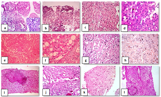
Photomicrographs: a) Hepatocellular carcinoma, trabecular pattern , inset shows intracytoplasmic inclusions.(H& E,x400). b) Hepatocellular carcinoma, clear cell variant (H&E, x100). c) Glycogen storage disease, hepatocytes with pale, glycogen rich cytoplasm (H&E,x400). d) PAS positive hepatocytes (PAS,x400 ). e & f) Liver Cirrhosis, regenerative nodules showing liver plates two or more cells thick.(H&E,x100,x400. g) Amyloidosis with amyloid deposited in the space of disse (H&E, x400). h) Congo red positive amyloid (Congo red stain, x400). i ) Miliary tuberculosis, inset shows granuloma (H&E, x100,x400). j) Biliary atresia showing ductular proliferation and neutrophilic infiltrate. (H&E, x400). k) Ballooning degeneration of liver (H&E, x400). l) Macrovesicular and microvesicular fatty change (H&E, x400).

Discussion
Patients who suffer from hepatosplenomegaly and present with an abnormal liver function test or unexplained jaundice, a liver biopsy is the best and the only way to attain the correct diagnosis. In our study ,out of 66 cases, 68.18% were males and 31.38% were females. The highest incidence of cases were reported in the sixth decade of life with preponderance in males. This is most probably due to higher incidence of alcohol usage by males than in females.4,5 The presenting symptoms were fever, nausea, vomiting, pain abdomen, decreased appetite and weight loss. Clinical findings included hepatomegaly, jaundice, pallor and ascites in varying combination in each case. Liver biopsy done for these cases yielded diagnosis in most cases while it showed non specific changes in 10.6 % of cases.
The most common histopathological diagnosis in the study was hepatocellular carcinoma (36.3%). This is in concordance with findings of Chawla et al (2013)4 but in contrast with Abdo et al (2006)6 and Ugaigbe et al (2013),7 who detected higher incidence of chronic hepatitis (Hepatitis C) than hepatocellular carcinoma. Hepatocellular carcinoma is one of the most common and devastating malignant tumors worldwide with variable geographic incidence due to differences in the major risk factors. It is usually a complication of chronic liver disease that is mainly caused by hepatitis B and/or C viral infections. Other important etiological risk factors are aflatoxin B1, alcohol, and cigarette smoking.7,8 Out of 24 cases of hepatocellular carcinoma, history of alcohol abuse was found in 58.33% (14 cases) and 20.83%(5 cases). Were positive for hepatitis B surface antigen(HBsAg). This is in concordance with the findings of Jhala et al.(2007)9 who found an association of hepatitis B surface antigen (HBsAg) with hepatocellular carcinoma in 27% cases seen in India. Histologically, in all cases, tumor cells were arranged in sheets, pseudoacinar pattern, trabecular pattern and/or cords having abundant eosinophilic cytoplasm and pleomorphic, hyperchromatic nuclei showing intranuclear vacuolations.(Figure 1).
Glycogen storage disease observed in 19.69% of the cases , was found to be the next common lesion. Koshy et al (2006)10 reported 17 cases from Southern India & indicated it may not be rare in India. Inherited disorders of glycogen metabolism affect the liver, muscle, heart and other systems. These disorders predominantly affect infants and children. The glycogen storage disease types that commonly involve the liver are types I, III, VI and IX. The distinction between these types is best done by enzyme studies of liver tissue obtained by wedge biopsy. Proper and early diagnosis of the glycogen storage disease is essential as dietary treatment with raw cornstarch and frequent high-starch feeds can prevent progression of the disease.10,11
Liver cirrhosis and steatosis were the third most common lesion seen in 4.54 %, probably attributable to increased alcohol intake or viral hepatitis. This incidence was close to Gall et al (1960) who studied liver biopsies over 25 years and found average in cidence of cirrhosis to be 6 %.4 Other rare diagnosis such as amyloidosis, biliary atresia, miliary tuberculosis etc should be kept in mind during the investigation of a liver disorder.
Conclusion
Microscopic examination of liver biopsy yields a diverse range of pathological findings. It is most important investigation in reaching accurate diagnosis, detect cause & severity of liver diseases and in providing better treatment options. Considering the value and safety of liver biopsy procedure, and the current limitations of noninvasive tests, liver biopsy will continue to remain as the cornerstone and the gold standard test in the assessment of liver diseases.
