Introduction
Galactoceles are common benign cystic lesions of breast.1, 2, 3 These can be seen often during pregnancy and lactation period. It has been noted that if such swelling remain over a period, then crystal formation may occur in these cystic lesions.2, 3 Sometimes the swelling can become so firm to hard that it can mimic malignancy on clinical and radiological evaluation.
Herein we report a rare case of Crystallizing Galactocele in a 25-year-old female who developed this swelling during her later part of pregnancy. The present case justifies the role of minimally invasive procedure like FNAC which is also simple, fast and economical.
Case Presentation
A 25-year-old female presented with swelling in the right breast for last 2 years. She noticed this swelling during her third trimester of pregnancy which has gradually increased in size over the period of last 2 years. The patient had this swelling during her pregnancy and lactational period. After delivery, she had been lactating since then. There was no history of trauma, or any kind of serous/bloody discharge from the nipple. On physical examination, a round to oval swelling was noted in the right breast in upper and outer quadrant. The swelling was mobile, non-tender, with smooth margin and no fixity to overlying skin and underlying chest. Nipple and areola were unremarkable. A clinical diagnosis of fibroadenoma was made.
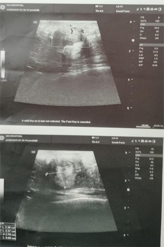
The patient was advised for ultrasound breast and fine needle aspiration cytology from the swelling in the right breast. Ultrasound showed a well-defined heterogenously hypoechoic to hyperechoic lesion of size measuring 2.3 x 2.2 cms in right breast at 10’o’clock position, 5-6 cm away nipple-areola complex with minimal vascularity [BIRADS-III]. A possibility of Fibroadenoma was given.(Figure 1)
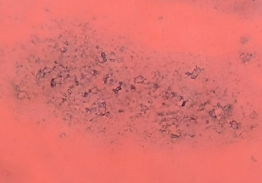
Figure 3
Smear showing group of benign ductal epithelial cells along with variably shaped crystals in background [Pap, 200X].
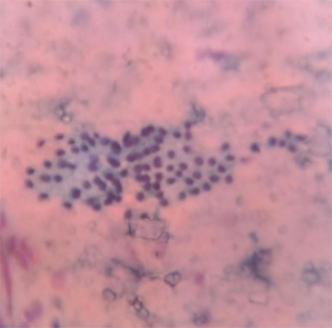
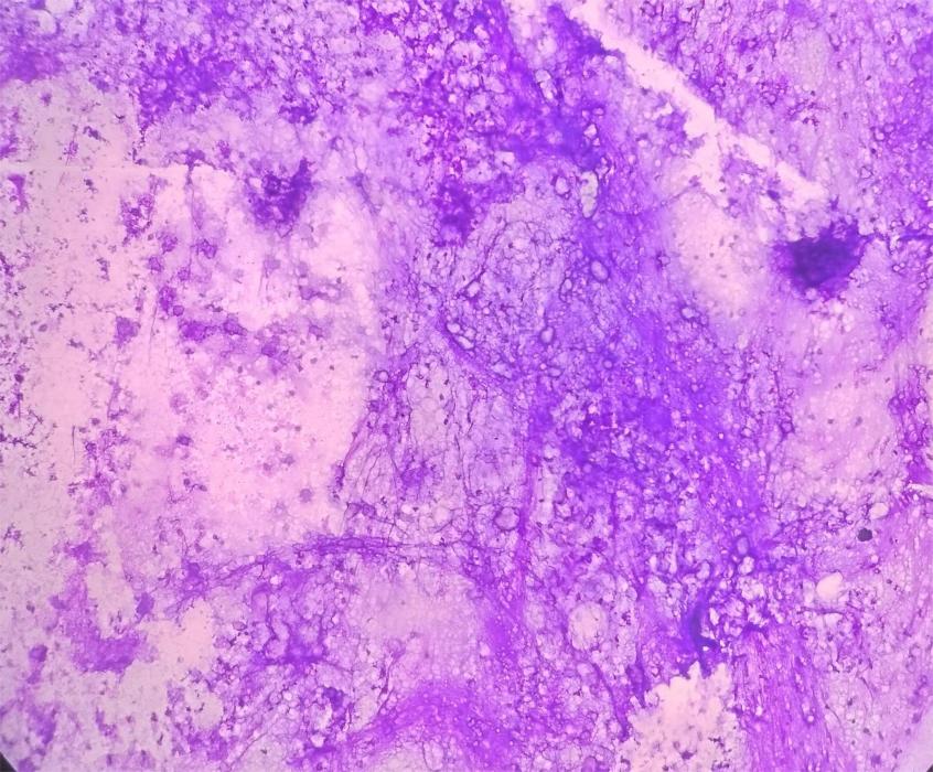
Figure 4
Smear showing amorphous granular proteinaceous material with frothy appearing micelles [Giemsa, 200X].
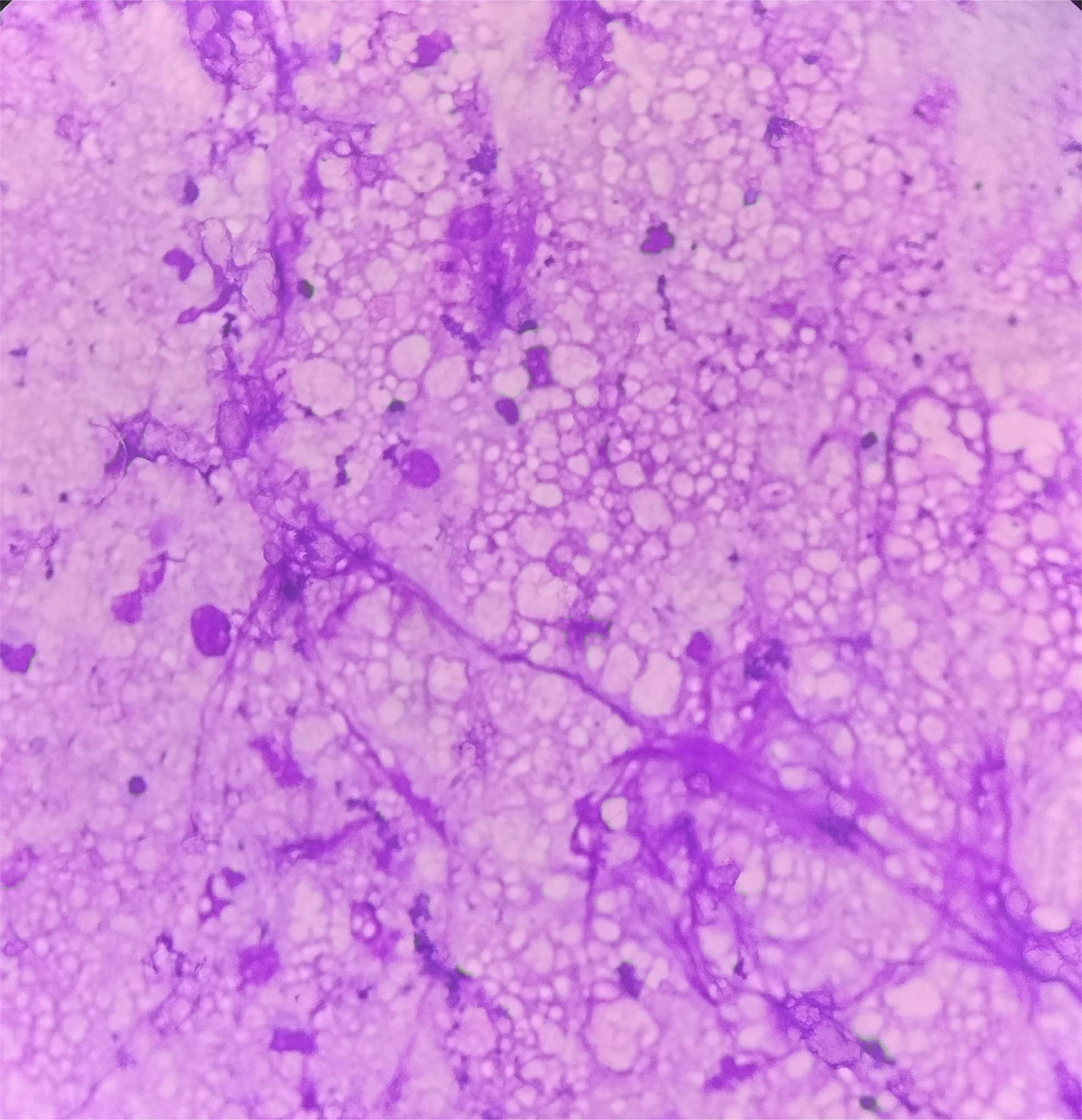
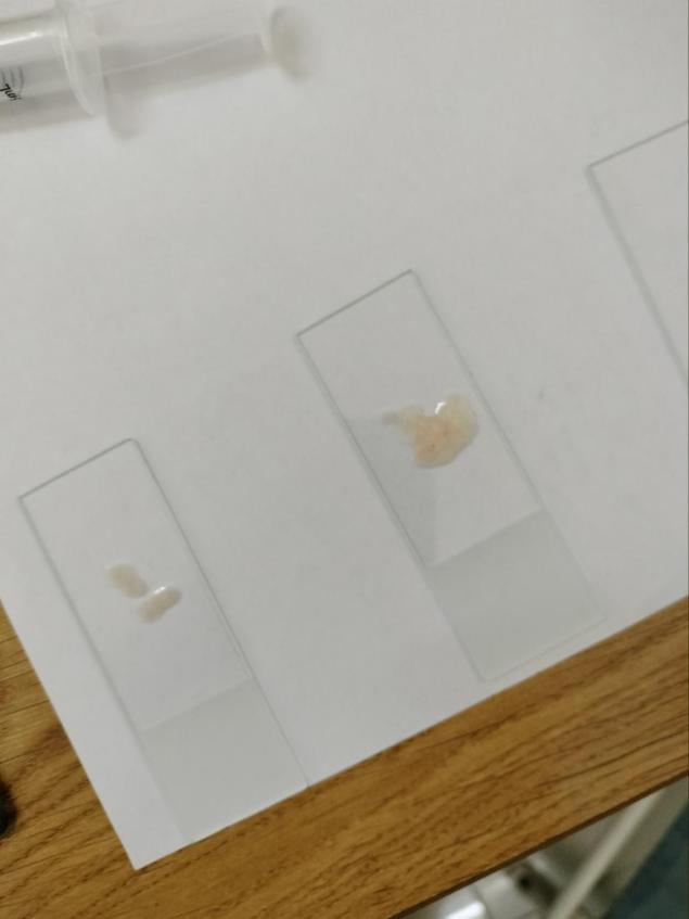
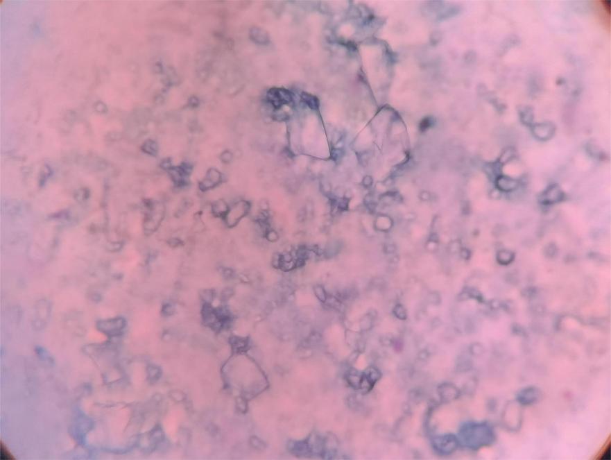
Fine needle aspiration yielded milky white fluid with whitish flake like material(Figure 2). Smears were stained with Giemsa and Papanicolaou stain. Smears examined showed occasional groups of benign ductal epithelial cells along with few scattered bipolar nuclei ($Figure 3. Background showed many macrophages along with amorphous granular proteinaceous material and frothy appearing micelles($)Figure 4, Figure 5 Smears also showed angulated and oval shaped crystals along with foci of fibromyxoid stroma($).Figure 6, Figure 7Based on a detailed cytomorphological findings, a diagnosis of Crystallizing Galactocele was given.
Discussion
Swelling in the breast is a common finding in females in all age groups.2, 3, 4 There are certain benign conditions which at times may mimic malignancy as well. These could be due to some internal physiological and pathological changes in the swelling. Such is a condition, termed as Galactocele of breast.2, 3, 4 It is a benign cystic lesion, more frequently noted in pregnancy and lactation, as was noted in our patient. Why this entity is usually found in these groups of female? These could be numerous factors like, stimulation by prolactin during pregnancy, because of secretory breast epithelium, obstruction of lactiferous ducts, etc.3, 4, 5, 6 Rarely it can be noted in nulliparous women with features of prolactinaemia and also in post-menopausal women. 3, 4, 5, 6, 7
Galactocele is basically a cystic collection of milk products that is lined by flattened cuboidal epithelium. Clinically, it presents as solitary, freely mobile lumps, usually in subareolar region. Due to physiological changes occuring in breast during pregnancy and lactation, its diagnosis becomes a challenging task.2, 3, 4, 5, 6, 7 Clinically, the lump gives an impression of fibroadenoma or lactating adenoma. Radiologically, depending on its contents, it can resemble an abscess, fibroadenoma, lactating adenoma or pregnancy-associated breast cancer.4, 5, 6, 7, 8 Hence, a pathological diagnosis is always needed to confirm the diagnosis for an early intervention and waive off the undue tension in the patient.6, 7, 8 With a proper pathological evaluation; using a very simple, minimally invasive and cost-effective diagnostic tool, i.e., FNAC, these entities can be rightly diagnosed.
Now comes the rare finding in the fact that, when these galactocele become long-standing, crystallization can occur in these galactoceles. Very few cases of crystallizing galactocele of breast have been reported in litertaure on FNAC.2, 3, 4, 5, 6, 7, 8, 9, 10, 11 In all cases no definitive opinion could be given on clinical and radiological grounds.4, 5, 6, 7, 8, 9, 10, 11 Milky fluid acts as a bed for these crsytal formation in long-standing cases. These secretions are composed of acid and neutral mucins, lipids, lysozyme, albumin, IgA, etc. Under acidic environment, supersaturation of these substances can land up in formation of tyrosine or calcium crystals. Gradually they become more firm to hard and at times they can mimic malignancy as well. 9, 10, 11 Hence timely diagnosis is warranted.
Grossly, it is a collection of inspissated lactational secretions, mainly fat and proteins. On cytological evaluation, we see dense amorphous proteinaceous material along with oval to angulated crystals of varying sizes and shapes. These crystals show birefringence on polarising microscopy. Special stains like per-iodic acid Schiff and von Kossa technique can be used to highlight these crystals. 4, 11
The course and outcome of crystallizing galactocele is benign and smooth without requiring major surgical intervention. They resolve of their own, sometimes therapeutic aspiration may be required depending the degree of symptoms and internal crystallization.4, 5, 6, 7, 8, 9, 10, 11 We report this condition to highlight the detailed cytological fetaures of crystallizing galactocele and also advocating to keep it as an important differential diagnosis while evaluating a breast mass in pregnant & lactational age group.
Conclusion
To conclude, during evaluation of breast swelling in pregnant and lactating females, differential diagnosis of crystallizing galactocele should always be kept. A simple, minimally invasive diagnostic tool can help in making a diagnosis of this rare phenomenon, thereby waiving off unnecessary surgical intervention in the patient.
Key points
Crystallizing galactocele of breast(CGB) should be considered an important differentials in evaluation of breast mass in pregnant and lactating females.
Only clinical and radiological findings are not sufficient to arrive at a definitive diagnosis.
Pathological diagnosis is mandatory.
Course and management of CGB is smooth without surgical intervention in majority of cases.

