- Visibility 79 Views
- Downloads 18 Downloads
- DOI 10.18231/j.jdpo.2022.066
-
CrossMark
- Citation
Hibernoma-A rare tumor on rare site
- Author Details:
-
Manika Alexander
-
Himachal Mishra *
Introduction
Hibernoma is a rare benign tumor which originates from brown fat. The tumour was first described as “pseudolipoma” by Merkel in 1906. It was named as hibernoma by Gery in 1914 because of its similarity to brown fat found in hibernating animals. Hibernomas accounts for <2% benign lipomatous tumors and 1% of all adipocytic tumors. It is seen in the middle-aged adults with equal sex distribution. It is thought to be arise from areas containing residual brown fat like- intrascapular area, axilla, neck, chest, abdominal cavity and retroperitoneum.[1] Tumor shows a predilection for subcutaneous tissue of the thigh, upper trunk, and neck.[2] Occasionally, hibernomas also seen to occur in sites in which brown fat is not found normally- extremities and limb girdles. Hibernomas which are large and deep-seated occuring in these sites cause diagnostic problems. Size of tumor vary from 1 to 24 cm with an average tumor size of 9.3 cm. Deep seated hibernomas accounts for 15%.[2]
Hibernomas composed 3 cell types found to occur in varying proportion of brown fat cells which are large with finely vacuolated cells having eosinophilic granular cytoplasm along with microvacuolated lipoblast-like cells and mature adipocytes.
It is not common to find tumors with predominantly multivacuolated lipoblast-like cells, but it is distinct from atypical lipomatous tumor. It is a benign tumor with neither any history of recurrence after a complete surgical excision nor any no case metastases to get reported till now. It has been found to show a characteristic finding of 4 11q13 rearrangement which results in MEN1 and AIP codeletion. [2]
Case Details
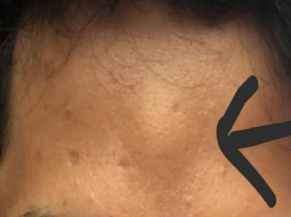
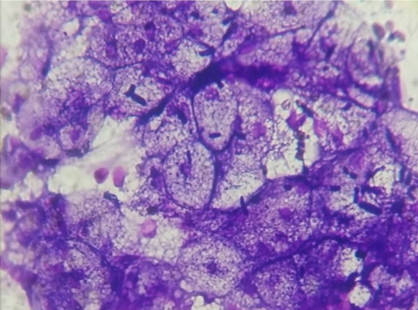
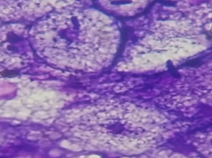
A 23 year old male presented with complain of a painless non progressive swelling over forehead for 6 months. Swelling was found to be 2x2cm cm, soft, mobile, non-tender, non-pulsatile with no skin changes. Ultrasonography of forehead swelling reported as Hyperechoic lesion measures 19 x 10 mm seen in subcutaneous plane- may be Lipoma. FNAC was performed. Microscopy shows sheets and clusters of partly or principally of coarsely multivacuolated fat cells with small central nuclei and no atypia; suggestive of Hibernoma- typical type.


Follow up: Since last 1 year, tumour is still non-progressive with no pressure symptoms and hence not underwent for surgery.
Discussion
Hibernoma tumor may occur rarely in forehead area and a few incidence over forehead has been reported. [3] Study done by Furlong et al, on 170 cases which reported to have most common sites of occurrence of hibernoma in body and includes-thigh, shoulder, back, neck, chest, arm, and abdominal cavity/retroperitoneum. [3] Even after multi diagnostic Imaging, radiological diagnosis remains difficult.[1]
The problem which arise to decide in preoperative stage to establish the diagnosis can be compounded with the help of radiological studies which shows a inhomogenous lipomatous tumor displaying prominent contrast enhancement. [4] A typical hibernoma contains a small bland fat droplets which is found to have occasionally finely granular and a small nucleus which is centrally located having a smooth contours with a evenly distributed chromatin. It is also recognised by another distinguishing features which aids in the cytological diagnosis by presence of delicate capillaries which surrounds a hibernoma cell.
Cytologically one can differentiate hibernoma from well differentiated liposarcoma based on the different appearance of brown fat-like cells in hibernoma and lipoblasts of liposarcoma. A well differentiated liposarcoma shows a varying size univacoulated or multivacoulated fat cells with an enlarged hyperchromatic nuclei showing scalloping or indentation by fat cells. [4] While a multivacoulated hibernoma cells often be confused for liposarcoma with multivacoulated lipoblasts. Nuclei of hibernoma lacks significant atypia and scalloping. Nuclear enlargement with mild atypia is seen in typical lipomatous tumors/well differentiated liposarcoma.[5]
Apart from well differentiated liposarcoma, a hibernoma must be differentiated cytologically from polygonal cell tumors which includes- Lipoma, Chondroid Lipoma, Myxoid Liposarcoma, Adult Rhabdomyoma, Atypical Lipomatous Tumor/Well Differentiated Liposarcoma, Paraganglioma and Alveolar Soft Part Sarcoma.
In case of lipoma with univacoular cell dominance, it becomes difficult to differentiate it from hibernoma. In such cases, presence of multivacoulated or granular cell favours to the diagnosis of hibernoma.[5] Another tumor which must be kept as differential is chondroid lipoma, which is a benign but a rare tumor having biphasic features of lipomatous component with small hibernoma-like cells, lipoblasts, and mature fat cells with variable chodromyxoid to hyalinized matrix.[4] Arborizing chicken wire like capillary vasculature is the characteristics features of myxoid liposarcomas.
Paraganglioma shows moderate degree of nuclear pleomorphism with occasional gland or follicular pattern and may resemble hibernoma. Alveolar arrangement with marked nuclear pleomorphism and prominent nucleoli are distinguishing features of Alveolar soft sarcoma.[5] To arrive at a correct diagnosis, one should weigh and assess clinical findings along with all the morphological features. Cytology of hibernoma often need to be differentiated from myxoid/round cell liposarcoma. A diagnosis of myxoid liposarcoma is favoured by presence of multivacoulated lipoblasts in a prominent myxoid background. Among the important cytological features, nuclear atypia is a diagnostic feature of round cell liposarcoma. Hibernoma showing finely granular cytoplasm can resemble benign granular tumor. Granular cell tumor can be distinguish from hibernoma by absence of mature lipomatous component, typical multivacoulated brown fat-like cells and a delicate vasculature. Cytologically, hibernoma should also be differentiated from adult rhabdomyoma which are large cells having dense fibrillary cytoplasm in absence of brown fat like cells. Electron microscopy further helpful in diagnosis of hibernoma. Microscopic features of hibernoma seen in electron microscopy includes- brown fat in the small vacuolated cells, small uniformly sized lipid droplets and numerous polymorphous mitochondria having thick inclusions with a close relationship with capillaries.[4]
In our opinion a reliable diagnosis of hibernoma can be made by combination of clinicoradiographic characteristics and unique cytologic features. But the cytopathologist should be aware of the diagnostic pitfalls leading to misdiagnosis of liposarcoma and benign fatty lesions with regressive changes.[4]
Conclusion
It is a small, benign, lobulated, non-tender lesion which occurs in adults with a peak incidence in the third decade. [6] It not be confused with atypical lipomas or well-differentiated liposarcoma.
Source of Funding
None.
Conflict of Interest
None Declared.
References
- D Daubner, S Spieth, J Pablik, K Zöphel, T Paulus, M Laniado. Hibernoma- 2 patients with a rare lipoid soft-tissue tumour. BMC Med Imaging 2015. [Google Scholar] [Crossref]
- AY Hmada, IM Schaefer, CDM Fletcher. Hibernoma Mimicking Atypical Lipomatous Tumor: 64 Cases of a Morphologically Distinct Subset. Am J Surg Pathol 2018. [Google Scholar] [Crossref]
- MA Furlong, JCF Smith, M Miettinen. The Morphologic Spectrum of Hibernoma- A Clinicopathologic Study of 170 Cases. Am J Surg Pathol 2001. [Google Scholar] [Crossref]
- MM Lemos, LG Kindblom, JM Meis-Kindblom, F Remotti, W Ryd, B Gunterberg. Fine-Needle Aspiration Characteristics of Hibernoma. Cancer 2001. [Google Scholar] [Crossref]
- JH Choi, DS Shin. Imprint cytology of Hibernoma- A case report. Korean J Cytopathology 2008. [Google Scholar]
- C Daniel, BL Robert, KL Hurtubise, EA Johnson. Symptomatic Hibernoma: A Rare Soft Tissue Tumor. Hawai Int J Med Public Health 2012. [Google Scholar]
