Introduction
The most common exocrine malignancy involving pancreas is Ductal Adenocarcinoma. Undifferentiated carcinoma in pancreas is a malignant neoplasm in which a substantial proportion of tumour does not show a definitive direction of differentiation with extremely poor prognosis and includes anaplastic undifferentiated carcinoma, Sarcomatoid undifferentiated carcinoma and carcinosarcomas. The undifferentiated carcinoma with osteoclastic giant cells is a distinctive rare neoplasm involving pancreas showing characteristic histomorphological findings comprising of variable admixture of mononuclear histiocytic cells, non-neoplastic giant cells and neoplastic mononuclear cell component. This tumor has been shown to share genetic alterations with pancreatic ductal adenocarcinoma, clinically, it behave unpredictably with a substantial proportion of patient showing prolonged survival period. We present a case of this rare pancreatic tumour with brief literature review discussing the key pathologic features, immunophenotype, genetic profile and clinical behavior.
Case Report
A 50 years female presented with progressive jaundice over a period of 2 months with direct hyperbilirubinemia of 4.5mg/dl. Computed tomography revealed a well defined space occupying mass lesion in the head of pancreas measuring 4.5cmx3.0 cm. Serum CA19-9 levels were mildly elevated. Endoscopic retrograde cholangiopancreatography (ERCP) was performed and a Fine needle biopsy was performed along with biliary stenting.
Figure 1
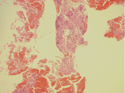
Hematoxylin and eosin stain. 10x. Low power view of lesion showing mononuclear cells admixed with osteoclastic giant cells.

Figure 2
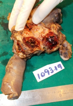
Whipple resection showing a well demarcated tumour in head of pancreas with visible hemorrahgic areas.

Figure 3
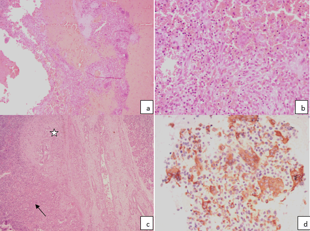
Hematoxylin and eosin stain; a: 10x showing hemorrhagic cystic areas lined by osteoclastic giant cells and admixed mononuclear cells; b: 40x Mononuclear cells exhibiting mild degree of nuclear pleomorphism with ovoid to spindled nuclei and admixed osteoclastic giant cells; c: Tumour area (arrow) with adjacent pancreatic duct showing low grade pancreatic intraepithelial neoplasia (star); d: CD68 immunostain highlighting osteoclastic giant cells and also some mononuclear cells.

Microscopy showed a tumor composed of intimate admixture of mononuclear cells along with osteoclastic type of giant cells and scant lymphomononuclear cells. The mononuclear cells were ovoid to spindled in shape with moderate amount of eosinophilic cytoplasm, dispersed nuclear chromatin and focal conspicuous nucleoli.(Figure 1) Mitosis were infrequent. In view of morphologic features, a possibility of Undifferentiated carcinoma with osteoclastic giant cells was suggested. Whipple resection was done and the excised specimen was subjected to histopathology. Gross examination shows a well- circumscribed and well-defined lesion in head of pancreas measuring 4.5x3.0x2.6cms, reddish brown in color with visible areas of hemorrhages and cystic spaces. (Figure 2) Microscopy showed variable sized hemorrhagic spaces lined by osteoclastic giant cells. (Figure 3a). The adjacent solid areas shows an infiltrative tumor with intimate admixture of mononuclear cells, osteoclastic giant cells (Figure 3b) communicating with pancreatic duct branches showing low grade pancreatic intraepithelial neoplasia changes (Figure 3c). Malignant glandular elements were not identified. Lymph nodes examined were uninvolved by tumour.
Immunohistochemistry was performed which showed immunopositivity of mononuclear cells for CD68 (Figure 3d). IHC for p53, Pan cytokeratin, CEA revealed negative results. Ki 67 index was 25-30%.
Discussion
Undifferentiated carcinoma of pancreas is a malignant pancreatic epithelial neoplasm in which a substantial component of tumor does not show a definitive evidence of phenotypic differentiation. It includes Anaplastic carcinoma, Sarcomatoid carcinoma, Carcinosarcoma and undifferentiated carcinoma with osteoclastic giant cells. 1
Undifferentiated carcinoma with osteoclastic giant cells is an extremely rare exocrine malignant neoplasm comprising less than 1% of all pancreatic malignancies.2 The age group involved is fifth to sixth decade with slight female predominance. Patients may present with varied symptoms such as nausea, fatigue, weight loss, tarry stools and jaundice. Serum tumor marker CA19-9 are elevated in majority of patients.3 Contrast enhanced Computed tomography scans is a sensitive modality to detect such lesions which shows mass lesions with slight enhancement of solid parts.4
Grossly, this tumor commonly involves body and tail of pancreas with a median size of 5cm, the largest reported tumor size being 24.5cm.5 Histologically, this tumor has classical morphologic appearance with admixture of non neoplastic osteoclastic giant cells, mononuclear histiocytic cells and neoplastic mononuclear cells. These neoplastic mononuclear cells may show varied cytologic features with spindled to epithelioid cells exhibiting variable nuclear hyperchromasia and pleomorphism. Additionally, malignant glandular component though not essential for diagnosis have been reported in variable percentage. Immunohistochemically, the histiocytic component - osteoclastic giant cells and mononuclear histiocytic cells are immunopositive for CD68 and Lyzozyme (marker of histiocytic differentiation). These cells are immunonegative for epithelial and mesenchymal markers. The neoplastic mononuclear component is variably positive for Vimentin and may or may not express keratin markers - CAM5.2, CK7 and CK19. p53 nuclear immunoreactivity is seen in majority of cases in malignant epithelial component. Associated pancreatic lesions, Intraductal pancreatic mucinous neoplasm3 and mucinous cystic neoplasm6 has been reported in few cases. Isolated cases with mantle cell lymphoma is also reported.
Since the epithelial component is not conspicuous in many cases, there has been varying opinions about the histogenesis of this tumor, however, there is enough evidence to suggest epithelial nature including co-existent malignant adenocarcinoma component in these tumors. Vimentin immunoexpression has been suggested to be due to mesenchymal transition from ductal cells.
Genetic study performed by Claudio et al7 in a series of 22 cases of undifferentiated carcinoma revealed shared molecular abnormality with pancreatic ductal carcinoma. Which includes alterations in KRAS tumour proto-oncogene and mutations in tumor suppressor genes- SMAD4, TP53, CDKN2A. These mutations comprise the commonly recurrent abnormality in pancreatic cancer genome constituting the four mountains in pancreatic cancer oncogenesis. Additionally, hotspot mutations in SERPINA 3(a member of serine protease inhibitor family) has been identified in a comprehensive genetic profile testing by Claudio et al.7 in two cases.
Surgical en block resection remains the first line of treatment. Despite sharing the genetic alterations with the conventional pancreatic ductal adenocarcinoma, the undifferentiated carcinoma with osteoclastic giant cells have dismal prognosis, though few long survivors are being reported. A meta-analysis carried out by Koboyashi et al.8 demonstrated that the different parameters are implicated in the short term survival includes older age, lymph node status positive, concomitant component of ductal adenocarcinoma. Our case did not show any malignant glandular component on histologic examination rather showed low grade pancreatic intraepithelial neoplasia and lymph nodes were uninvolved by tumour. He is on regular follow up with no evidence of recurrent disease or metastasis after 1.5 years of surgical excision.
Conclusion
Undifferentiated carcinoma with osteoclastic giant cells is a rare malignant neoplasm involving pancreas with distinct histomorphological appearance and shared molecular aberrations with pancreatic ductal Adenocarcinoma. These tumors are biologically distinct from ductal Adenocarcinoma of pancreas with better survival period.
