Introduction
Salivary glands are unique among the secretary glands with a more heterogenous group of tumors showing greatest histological diversity. Salivary gland system includes three pairs of major glands – Parotid, Submandibular and Sublingual and many minor glands in the mucosa of the oral cavity, lips, floor of mouth, gingival, cheek, hard and soft palate, tonsillar areas and oropharynx.1 The spectrum of salivary gland lesions is wide and the relative incidence of neoplastic versus non neoplastic lesions is variable in different studies.2 Salivary gland tumors account for 2% of all human neoplasm and relatively uncommon.3 Salivary glands tumors are rare with annual incidence <1/100000 inhabitants. These tumors show wide range of morphological diversity between different tumor types and sometimes within an individual tumor mass. 4 About 65% to 80% arise within the parotid, 10% in the submandibular gland and the remainder in the minor salivary glands including the sublingual gland.5 Tumors have the highest chance of malignancy if they arise from retromolar area(89.7%), floor of mouth(88.2%),tongue(85.7%)and sublingual gland(70.2%) whereas only 20% of all parotid tumors are malignant.6
The aim of the study is to recognize various histopathological patterns of salivary gland lesions, their frequency, age, gender and site wise distribution.
Materials and Methods
The present study “Histopathological study of salivary gland lesions” is carried out in the Department of Pathology, KBNIMS, Kalaburagi. It is a two year retrospective study from November 2018 to November 2020. The blocks were retrieved and sections were cut and stained with Haematoxylin and Eosin. Information regarding age, sex, site, complaints, clinical and radiological findings were recorded from requisitions received in the histopathological department. Special stain PAS was used when required. The tumors were classified into nonneoplastic and neoplastic lesions according to WHO histological typing of salivary gland tumors.
Results
Total 50 cases were included. Both gross specimens and biopsies which were received for histopathological examination, during the period of 2 years i.e from Nov 2018 to Nov 2020 were included. Out of these 50 cases, 27 cases (54%) were diagnosed as non-neoplastic lesions and 23 cases (46%) as neoplastic lesions.
Table 1
Age wise distribution of salivary gland lesions
|
Age group in years |
Percentage |
|
10-20 |
- |
|
21-30 |
03(6%) |
|
31-40 |
26(52%) |
|
41-50 |
09(18%) |
|
51-60 |
10(20%) |
|
61-70 |
02(4%) |
Most of the cases showed male predominance and were commonly found between the age group 31-40.
Table 3
Site wise distribution of salivary gland lesions
|
Paroti D Gland |
18(36%) |
|
Submandibular Gland |
27(54%) |
|
Minor Saliwary Gland |
05(10%) |
Most common site involved was submandibular region.
Table 4
Spectrum of non-neoplastic salivary gland lesions
|
Chronic Sialadenitis |
16(32%) |
|
Granulomatous Sialadenitis |
03(6%) |
|
Mucocele |
05(10%) |
|
Retention cyst |
03(6%) |
Chronic Sialadenitis was the common non neoplastic salivary gland lesion accounting for32%
Table 5
Spectrum of neoplastic salivary gland lesions
|
Pleomorphic Adenoma |
11(22%) |
|
Warthins tumor |
05(10%) |
|
Mucoepidermoid carcinoma |
03(6%) |
|
Adenoidcystic carcinoma |
02(4%) |
|
Epithelial myoepithelial carcinoma |
01(2%) |
|
Acinic cell carcinoma |
01(2%) |
Pleomorphic adenoma was the most common benign tumor and Mucoepidermoid carcinoma was the most common malignant tumor found in the present study.
One case of mucoepidermoid carcinoma was found in 24 year male in the submandibular region. Rare malignant tumor, Epithelialmyoepithelial carcinoma was diagnosed in 50 year male in the parotid gland.
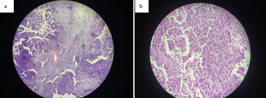
Figure 1
a: Pleomorphic adenoma composed of chondroid matrix. b: Pleomorphic adenoma composed of epithelial element comprising of tubular, ductal andsquamoid differentiation

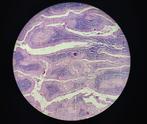
Figure 2
Warthin’s tumor: Blunt papillary projection exhibiting double layer of oncocytic lining cells and underlying lymphoid stroma.

Discussion
Total number of salivary gland lesions includes 50 cases. Predominantly the cases were found in the age group of 31-40 which was similar to Syed imtiyaz et al study.
Table 6
Comparative analysis of age wise distribution of salivary gland lesions
|
Age group |
Syed Imtiyaz Hussain et al 7 |
Richa study7 |
Present Study |
|
0-10 |
7% |
6.32% |
- |
|
11-20 |
8.86% |
- |
|
|
21-30 |
21.0% |
27.84% |
06% |
|
31-40 |
26.0% |
18.98% |
52% |
|
41-50 |
22.0% |
12.65% |
18% |
|
51-60 |
14.0% |
15.2% |
20% |
|
61-70 |
10.0% |
8.86% |
04% |
|
71-80 |
|
- |
|
|
81-90 |
1.26% |
- |
Table 7
Comparative analysis of salivary gland lesions in males and females.
|
Study |
M:F RATIO |
|
|
Benign |
Malignant |
|
|
Dave P.N. et al 8 |
1 |
1.42 |
|
Syed Imtiyaz Hussain et al |
1.62 |
4.28 |
|
Kirti N. Jaiswal et al 9 |
0.7 |
1.28 |
|
Present Study |
1.54 |
1.21 |
The present study showed male predominance in both benign and malignant salivary gland lesions which was similar to other above mentioned studies
Table 8
Comparative analysis of salivary gland lesions in different studies.
|
Characteristics |
No of Cases |
Percentage (%) |
||||
|
Non Neoplastic |
Benign |
Malignant |
Non Neoplastic |
Benign |
Malignant |
|
|
Mallepogu Anil Kumar et al 10 |
15 |
30 |
10 |
27.27% |
54.54% |
18.18% |
|
Dhanamjeya Rao Teeda et al 11 |
12 |
31 |
10 |
22.64% |
58.49% |
18.86% |
|
Malliga .S et al 12 |
21 |
53 |
29 |
20.4% |
51.45% |
28.15% |
|
Richa study 1 |
28 |
34 |
17 |
35.44% |
43.03% |
21.51% |
|
Present Study |
27 |
16 |
07 |
54% |
32% |
14% |
In the present study, non neoplastic lesions were 54% and it was more, compared to other above mentioned studies. Benign tumors were 32% and it was less, compared to other above mentioned studies. Malignant tumors were 14% and it was less, compared to above mentioned studies.
Table 9
Comparative analysis of site wise distribution of salivary gland lesions.
|
Site |
Dhanamjeya Rao Teeda et al 11 |
Anita Omhare et al 13 |
Mallepogu Anil Kumar et al 10 |
Malliga. S et Al 12 |
Present Study |
|
Parotid Gland |
73.5% |
48.3% |
67.27% |
58.25% |
36% |
|
Submandibular Gland |
16.9% |
41.2% |
25.45% |
29.12% |
54% |
|
Minor Salivary gland |
9.4% |
10.4% |
7.27% |
12.6% |
10% |
Most common site involved was Submandibular gland followed by Parotid and minor salivary gland in the present study while other studied showed Parotid gland as commonest site of involvement.
Table 10
Comparative analysis of non-neoplasic Salivary gland lesions
|
Type of Lesions |
Dhanamjeya Rao Teeda et al 11 |
Anita Omhare et al 13 |
Mallepogu Anil Kumar et al 10 |
Present study |
|
Acute Inflammation |
- |
- |
- |
|
|
Chronic Sialedenitis |
5.66% |
39.2% |
16.36% |
32% |
|
Tuberculosis |
- |
3.33% |
- |
6% |
|
Cystic Lesion |
16.98% |
9.2% |
10.9% |
- |
|
Rannula |
- |
- |
- |
- |
|
Retention Cyst |
- |
- |
- |
6% |
|
Mucocele |
- |
- |
- |
10% |
Among nonneoplastic lesions, most common lesion encountered was chronic sialadenitis and it was similar to Anita et al study.
Table 11
Comparative analysis of Benign Salivary gland tumor
|
Type of Tumors |
Dhanamjeya Rao Teeda et al 11 |
Anita Omhare et al 13 |
Mallepogu Anil Kumar et al 10 |
Malliga. S et al 12 |
Present Study |
|
Pleomorphic Adenoma |
45.25% |
21.66% |
43.63% |
41.6% |
22% |
|
Warthin’s Tumor |
5.66% |
0.8% |
10.9% |
2.1% |
10% |
|
Canalicular adenoma |
- |
- |
- |
- |
- |
|
Myoepithelioma |
1.88% |
- |
- |
- |
- |
|
Oncocyst adenoma |
- |
- |
- |
1% |
- |
|
Basal Cell Adenoma |
- |
- |
- |
- |
- |
|
Monomorphic Adenoma |
5.66% |
8.33% |
- |
- |
- |
|
Hemangioma |
- |
1.67% |
- |
- |
- |
|
Neurofibroma |
- |
- |
- |
1% |
- |
Among neoplastic lesions, most common benign lesion encountered was Pleomorphic adenoma and it was similar to Anita et al study.
Table 12
Comparative analysis of malignant Salivary gland tumor
|
Type of Tumors |
Dhanamjeya Rao Teeda et al 11 |
Anita Omhare et al 13 |
Mallepogu Anil Kumar et al 10 |
Malliga. S et al 12 |
Present study |
|
Mucoepidermoid Carcinoma |
9.43% |
6.66% |
7.27% |
22.9% |
06% |
|
Acinic cell carcinoma |
- |
3.33% |
- |
- |
02% |
|
Adenocystic Carcinoma |
3.77% |
1.66% |
3.63% |
4.2% |
04% |
|
Myoepithelial Carcinoma |
- |
- |
- |
- |
02% |
|
Salivary duct Adenocarcinoma |
1.88% |
0.8% |
- |
- |
- |
|
Carcinoam ex pleomorphic adenoma |
1.88% |
3.33% |
3.63% |
6.25% |
- |
|
Mammary analog secretory carcinoma |
- |
- |
- |
- |
- |
|
High grade B cell extranodal Non Hodgkins Lymphoma |
- |
- |
- |
- |
- |
|
Poorly differentiated Carcinoma |
1.88% |
- |
3.63% |
- |
- |
Among neoplastic lesions, most common malignant lesion encountered in the present study was Mucoepidermoid carcinoma and it was similar to Anita et al study. Epithelial myoepithelial carcinoma, a rare tumor was also found in our study.


