Introduction
Salivary gland lesions are not so common, especially neoplasms, which constitute less than 1% of all tumors and about 4% of all epithelial neoplasms encountered in the head an d neck region.1 These comprise a wide variety of benign and malignant neoplasms, non-neoplastic lesions which exhibit difference not only in biological behaviour but in prognosis as well. Tumours of salivary glands have continuously interested medical professionals, pathologists in particular because of a number of peculiarities of the subject. Approximately 80% of the salivary gland tumors are found in the Parotid gland and 10 to 15% in the submandibular gland. Majority of Salivary gland tumours are of benign histology (80-85%), with pleomorphic adenoma being the most common,(2) constituting 70% of benign tumours. The probability of malignancy is relatively inversely proportional to the size of the gland. Overall, benign tumors of the salivary glands tend to present somewhat earlier than malignant ones. The etiology, prognostic factor s and risk factors are poorly defined. Many of these lesions behave in an indolent fashion and some of the histologic types tend to recur late. Thus there is a call for follow up to improve the ability of the clinician to draw conclusion about the efficacy of treatment. Due to a lack of long term follow up, screening and registration, risk factors and prevention are poorly known.2 A diagnosis of salivary gland neoplasm must be considered in any patient who presents with a mass in the parotid or submandibular region or a sub mucosal mass in the oral cavity or pharynx. A preoperative sonography combined with FNAC, CT scan and MRI in some cases provides necessary clues prior to surgery.1 Although FNAC is a tool for preoperative evaluation, Histopathology still remains the gold standard in giving the final diagnosis.
Materials and Methods
Type of study
A retrospective and cross sectional study of the histopathological changes in biopsies of salivary gland lesions that were conducted in Department of Pathology, Government medical college, Kota from January 2015 to December 2017 (3 years).
Source of data
The present study titled “Histopathological spectrum of salivary gland lesions in Hadoti region” was conducted in Dept. of Pathology Govt. medical college, Kota from January 2015 to December 2017. The source of data is from the biopsies of lesions of salivary glands that were received at Department of Pathology from the Government hospital, Kota. A total of 138 cases were studied.
Methods of collection of data
The study was made according to the pre-designed proforma. The history and other details of clinical examination was taken from requisition form. The biopsies were subjected to routine tissue processing and stained with H&E, PAS & mucicarmine stain as per requirement..
Results/Observations
The present study included all the cases of salivary gland lesions that were reported in the Dept. of Pathology, Government Medical College Kota over a period of three years that is from January 2015 to December 2017. This study included a total of 138 cases.
Maximum number of cases were seen in Parotid gland constituting 81 cases (60%) followed by Submandibular gland constituting 45 cases (31%).
Table 1
| Parotid Gland | Submandibular Gland | Minor salivary Gland | Total | |
| Total | 81 | 45 | 12 | 138 |
| Percentage | 60 | 31 | 9 |
Showing site of lesion
Maximum number of non neoplastic lesions were common in 3rd to 4th decade, benign neoplastic in 4th to 5th decade and malignant neoplastic in 5th to 7th decade.
The above table shows male preponderance with M: F Ratio – 1.5:1.
Maximum numbers of salivary gland lesions were neoplastic- 75 cases (75.3%)
Table 3
| Non neoplastic | Neoplastic | Total | |
| No: of Cases | 63 | 75 | 138 |
| Percentage | 45.66 | 54.34 |
Nature of salivary gland lesions
Maximum numbers of salivary neoplasms are benign 59 cases (75.6%)
Table 4
| No: of Cases | Percentage | |
| Benign | 59 | 78.64 |
| Malignant | 16 | 21.36 |
| Total | 75 |
Incidence of salivary neoplasms
Common sites of all lesions were parotid (60%) followed by submandibular (31%) and minor salivary glands (9%) in order of frequency.
Table 5
Incidence of lesions
Chronic Sialadenitis is the most common lesion with 50 cases (36.28%) followed by Pleomorphic adenoma -49 cases (35.50%).
Table 6
Morphological spectrum of lesions
Figure 1
Adenoid cystic carcinoma: Showing adenoid and cystic components (H & E), showing neural involvement (x 400 X).

Figure 2
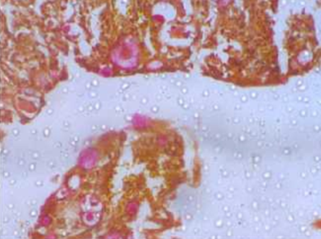
Mucoepidermoid Carcinoma: Showing Squamous and Mucinous Components (x !00X), with Mucicarmine positivity

Discussion
The salivary gland disorders represent a distinct group of disorders affecting both the major and minor glands. These conditions range from inflammatory disorders of infectious, granulomatous, auto immune in etiology to obstructive, developmental, idiopathic disorders and neoplasms.
Among the salivary lesions studied, maximum cases were neoplastic - 75 cases (54.45%) and the rest non-neoplastic cases 63(45.64%). Among the neoplasms studied, 59(78.64%) cases were benign and 16(21.36%) were malignant. This observation is comparable to most of the studies including case series, including those by Nepal et al, Ali NS et al, and Moghadam S A et al. where they noted a predominance of benign tumors over the malignant ones. Table shows that majority of the lesions were benign neoplasms, findings similar to other studies in literature.
Among the neoplastic lesions, maximum incidence was seen with benign neoplasms. Among these neoplasms, pleomorphic adenoma was most frequent, followed by Basal cell adenoma and Warthin’s tumor. Muco-epidermoid carcinoma was the most common malignancy observed.
In our study of 138 lesions, among the neoplastic lesions pleomorphic adenoma was the commonest, while among non-neoplastic lesions commonest was chronic non specific sialadenitis. Out of 63 non neoplastic cases, there were 49 chronic non specific sialadenitis (77.77%) & 6 mucus retention cysts.(9%). Majority of cystic lesions occurred in submandibular gland. In our study, it is noted that non neoplastic lesions were common in the 3rd and 4th decade of life. Benign tumors were common in 3rd to 5th decade and malignant tumors were common from 5th decade onwards
Table 7
Shrestha S et al. (2014)3 did a retrospective study of 176 cases of salivary gland tumors at Koirala Hospital, Nepal. The mean age observed was 44.76 years with age range of 12-75 years. Pleomorphic adenoma was found to be the commonest benign tumor (72.7%), followed by Warthin tumor (15.1%), monomorphic adenoma (3.0%) and basal cell adenoma (3.0%). Dr. Shazia Bashir et al conducted a combination study with retrospective data of 8 years and prospective data of two years
Out of total 80 cases, 49(61.25%) were benign and 31(38.75%) were malignant. Predominance of males was observed with M: F ratio of 2.3:1. The mean age observed was 44.76 years with age range of 12 to 75 years. Benign tumors outnumbered the malignant ones. Parotid was the most common site for the location of tumors (65%) followed by submandibular (25%) and minor salivary glands (10%). This is in conformity with other workers, viz., Gore et al.5 Richardson et al6 and Dandapat et al.7 Pleomorphic adenoma was the commonest salivary gland tumor observed in both sexes. Mucoepidermoid carcinoma was the most common among the malignant salivary gland tumor followed by adenoid cystic carcinoma. The sex incidence varied with respect to different lesions of the salivary glands. In the present study there were 24 males (45.28%) and 29 females (54.71%), with a M : F ratio of 0.8:1. Dandapat et al.7 and Rewsuwan et al.8 also reported a female preponderance in their series. Among the lesions of parotid gland majority were benign tumours (66.03%). Pleomorphic adenoma was the most common lesion with 28 cases (52.83%).
Table 8
| Bashir. S. et al (2013)4 | Erik G. Cohen et al (2004) | T.Chatter jee et al (2000)9 | Present study | |
| Parotid gland | 65.30% | 74 | 77 | 60% |
| Submandibular gland | 20.40 | 26 | 9 | 31% |
| Minor gland | 4.08 | - | 14 | 9.0% |
Comparision of Sit e of Salivary Lesions
The above table shows that Parotid gland is the frequent site in the present series among the salivary glands to have lesions as compared to the similar studies in literature. T. Chatterjee et al conducted a retrospective study for 23 years and 315 salivary gland specimens were received. There were 192 (61%) benign neoplasms and 123 (39%) malignancies. Among the benign ones, Pleomorphic adenoma was common and among malignancies, Adenoid cystic carcinoma was common. Parotid gland is the frequent site followed by Submandibular gland. Maximum Benign cases were seen in the third decade and malignancies in the 5th decade. Out of 53 cases studied there were 24 cases of pleomorphic adenoma, 9 cases were of cysts, 5 cases were mucoepidermoid carcinoma, 3 cases were monomorphic adenoma and Warthin’s tumour, 2 cases of Adenoid cystic carcinoma. There was 1 case each of myoepithelioma, salivary duct carcinoma, poorly differentiated carcinoma, carcinoma ex Pleomorphic adenoma. Out of 9 cysts received 3 cases were seen in the parotid gland, 1 case was in the submandibular gland, and 5 cases were in the minor salivary gland. Most of the cysts were of mucus retention type and mostly occurred in the minor salivary glands. Those in the major salivary glands were salivary duct cysts and retention cysts. In the present study, mean age for the occurrence of cysts was 41.1 years with age range of 12 to 70 years. There were 41 cases of salivary tumors, out of which 31 cases were benign tumor sand 10 cases were malignant tumors
Table 7 shows that pleomorphic adenoma is the most frequently encountered neoplasm of the salivary glands in the present study and is comparable to the similar studies by Sreshta et al, Erik G. Cohen et al, Naeem Sultan Ali. According to Foote and Frazell (1954)10 and G.G.Potdar,9 65 to 75 % of the tumours were pleomorphic adenomas. 24 cases (45.28%) encountered in parotid, submandibular and minor salivary glands. Most of these cases occurred in the parotid gland (82.35%). Potdar and Paymaster9 reported 183 cases of pleomorphic adenomas, out of which 101 were involving parotid gland. In the present study, mean age for pleomorphic adenoma was 35.53 years with age range of 20 to 65 years. Out of all reported cases of pleomorphic a denoma, 22 were males and 27 were females with a male to female ratio of 1:1.23.
There were 16 cases of malignant salivary neoplasms, out of which 6 were Mucoepidermoid carcinoma, 3 each of Adenoid cystic carcinoma and squamous cell carcinoma, 2 cases each of Salivary duct carcinoma and Acinic cell carcinoma. Mucoepidermoid carcinoma was the most common malignant salivary tumor occurring in the salivary glands, constituting 6(37.50%) of all malignant salivary gland tumors in the present study. Mucoepidermoid carcinoma was reported as the most common malignant salivary gland tumor of parotid by Richardson et al6 and Ali et al11. There were 3 cases of Adenoid cystic carcinoma. It is the second most common malignancy of the salivary glands in the present study. Findings corroborating with the series reported by Vergas et al12. In contrast to the present study, Lima et al and Rewsuwan et al8 reported adenoid cystic carcinoma to be the most common malignant salivary gland tumor in their series.
Conclusion
The Present study included lesions of salivary glands, presented in the Department of Pathology, Government Medical College, Kota from January 2015 to December 2017. Approach was to study the various histopathological types of salivary gland lesions, their classification and thorough study of lesions of salivary glands and to compare the observed findings to similar studies in relation to incidence, age, sex and risk factor distribution.
Following observations were noted
During the specified period, total of 138 cases of salivary gland lesions were studied.
Mean age observed was 32.7 years with an age range of 11 to 70 years.
There were 84 males (60.86%) and 54 females (39.13%), with a Male : Female ratio of 1.5: 1.
Parotid was the commonest site of lesion (73.5% of all tumors and 60% of all lesions)
Maximum cases were neoplasms - 75 cases (54.35%) and the non-neoplastic cases were 63(45.66%).
Majority of cases among non-neoplastic lesions were chronic non specific sialadenitis.
Among the neoplasms studied, 59(78.64%) cases were benign and 16 (21.36%) were malignant.
Pleomorphic adenoma was the most frequent histological type of benign neoplasm.
