- Visibility 204 Views
- Downloads 35 Downloads
- Permissions
- DOI 10.18231/j.jdpo.2019.057
-
CrossMark
- Citation
Frequency of HER2/neu over expression in adenocarcinoma of the gastrointestinal system, a Study of 70 cases
- Author Details:
-
Mansi Mukesh Thacker *
-
Sneha Chiplonkar
Abstract
Aim: To study the frequency of HER2/neu overexpression in esophageal, gastric, small intestine and colorectal adenocarcinomas, to study the association of HER2/ neu overexpression with histopathological differentiation of esophagus, stomach, small intestine and colorectal adenocarcinomas and identifying its significance.
Materials and Methods: Cross sectional study was performed on 70 cases which include resected specimens of stomach, small intestine and colorectal adenocarcinoma and biopsies of esophagus and these organs. The samples were divided into Group A (Esophagus and stomach) and Group B (Small intestine and colorectal). The samples were grossed and processed in the pathology department, and sections were stained with hematoxylin and eosin stain for histopathological confirmation of malignancy and graded as well-differentiated (gradeI), moderately-differentiated (grade II) and poorly-differentiated (gradeIII ) adenocarcinomas. The confirmed samples were processed for immunomarker study of HER2/ neu.
Results: Over expression of HER2/ neu was found in 17(24.3 %) patients overall. Out of 17 HER2/ neu positive cases, 4(26.7%) were from group a, while 13(23.6%) were from group b. No staining or membrane staining in < 10% tumor cells was observed in 46(65.7%) patients, which were labeled as score “0” and considered negative for HER2/ neu overexpression. Faint/barely perceptible membranous staining in ≥ 10% of tumors cells were observed in 5 (7.1 %) patie nts, which were labeled as score “1” and considered negative. Weak to moderate complete, basolateral or lateral membranous staining in ≥ 10% tumor cells were observed in 2(2.9%) patients, which were labeled as score “2” and considered equivocal. Strong complete, basolateral or lateral membranous staining in ≥ 10% of tumors cells were observed in 17(24.3%) patients, which were labeled as score “3” and considered positive. Out of 70 patients, 37(52.9%) were suffering from grade-Ⅱ malignancy, 17 (24.3 %) from grade- I and 16 (22.8%) from grade-Ⅲ. According to histological grades, frequency of HER2/neu over expression was 11.8% in well differentiated adenocarcinoma (grade I), 29.7% in moderately differentiated adenocarcinoma (grade II) and 25% in poorly differentiated adenocarcinoma (grade III). There was no statistical significance of HER2/neu expression and grades of tumor.
Conclusion: HER2/neu is over expressed in adenocarcinoma of gastroesophageal junction and colorectal adenocarcinoma. There is no statistical significance of HER2/neu expression and grades of tumor.
Introduction
HER2/neu oncogen is one of the four epidermal growth factor receptors. It is located on chromosome 17q21 and encodes a 185 transmembrane protein with tyrosine kinase activity which functions as a growth factor receptor.[1] It plays an important role in cellular proliferation and differentiation during fetal development. Gastric cancer is the fifth most commonly diagnosed cancer and the third most common cause of cancer related death globally[2] and colorectal carcinoma is the third most commonly diagnosed cancer in both men and women.[3] Amplification of the gene and over expression of this protein is seen in various malignancies other than gastrointestinal system such as cancers of breast,[4] lungs, prostate, bladder, pancreas, and in osteosarcoma.
The rate of esophageal, gastric, small intestinal and colorectal adenocarcinoma is increasing in India. Surgical resection is the mainstay of therapy for these carcinomas. Most gastric and esophageal cancers are diagnosed in a more advanced or unresectable stage; newer therapeutic strategies, treatment options and novel therapeutic targets are desperately needed.
HER2/neu is important as a target of the monoclonal antibody trastuzumab (marketed as Herceptin). Trastuzumab is effective only in cancers where the HER2/ neu receptor is overepressed. The mechanism of trastuzumab is that it binds to HER2/ neu and increases p27, a protein which halts cell proliferation.[5] Recently, the EMEA (European Medicine Agency) has approved use of trastuzumab in combination with chemotherapy in patients with advanced gastric cancer.[6],[7]
HER2/neu over expression in esophageal adenocarcinoma is associated with increasing depth of invasion, lymph node and distant metastases and poor survival.[8] The data available in the literature for HER2/neu positivity rates in gastric cancers vary from about 7%-43%. HER2/ neu positive status in gastric cancer also appears to be associated with more aggressive disease, a poorer prognosis, and shorter survival.
The purpose of the study is to document frequency of HER2/neu overexpression in esophagus, stomach, small intestine and colorectal adenocarcinomas and also to evaluate possible association of this parameter with histopathological differentiation and identifying its significance.
Materials and Methods
This study was cross sectional study performed on resected specimens of stomach, small intestine and colorectal adenocarcinoma and biopsies of esophagus and these organs submitted to the department of pathology during the period of two years. A total of 70 cases were studied. Clinical details of patient were obtained from requisition form received in pathology department and entry was made in the proforma.
The specimens were subjected to a thorough gross examination. Appropriate sections were taken. The sections were processed and paraffin blocks were prepared. The sections were cut at 4-5 micron thickness and stained with hematoxylin and eosin for histopathological confirmation of malignancy. The histological grading of these tumors was done as well-differentiated (I), moderately differentiated (II), and poorly-differentiated (III) according to the criterion of the American Joint Committee on Cancer. The confirmed malignant specimens and biopsies were processed for immunomarker study of HER2/ neu. Known positive breast cancer cases were used as positive controls, with the omission of primary antibodies for the negative control. For immunohistochemical staining by HER2/ neu, m onoclonal antibody of C-erbB-2 gene product was used (clone TAB250), supplied in a liquid form ready to use.
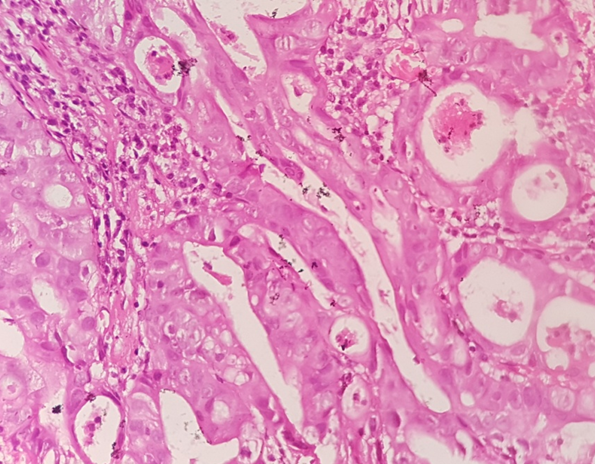
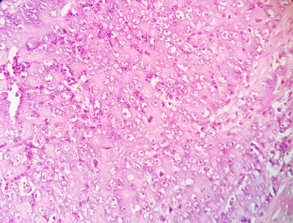
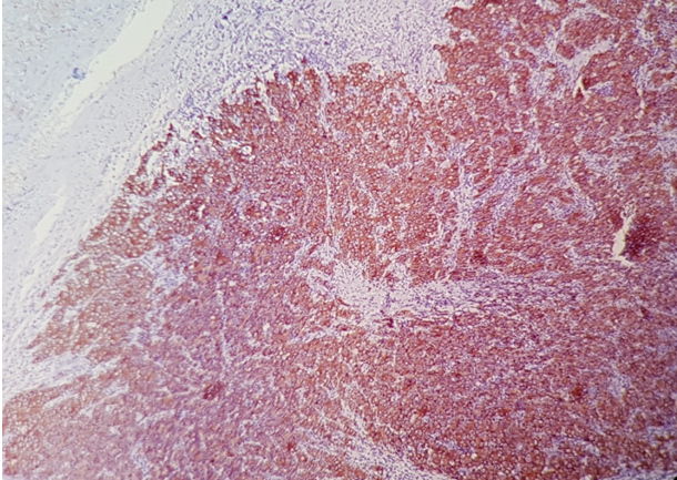
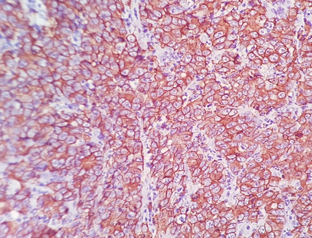
The Immunohistochemistry slides were scored according to scoring system by Hoffman et al.[9] No staining or membrane staining in < 10% tumor cells ,were labelled as score “0” and considered negative for HER2/neu overexpression. Faint/barely perceptible membranous staining in ≥ 10% of tumors cells were labelled as score “1” and considered negative. Weak to moderate complete, basolateral or lateral membranous staining in ≥ 10% tumor cells were labelled as score “2” and considered equivocal. Strong complete, basolateral or lateral membranous staining in ≥ 10% of tumors cells were labelled as score “3” and considered positive.
Study group were divided in Group A and Group B. Group A includes adenocarcinoma of gastro esophageal junction and gastric carcinoma and Group B includes small intestine and colorectal adenocarcinoma. Frequency of HER2/neu over expression in these study groups was evaluated and its association with histopathological grading was estimated. The data analysis was entered in MS-Excel sheet & was done by descriptive statistics for frequency/percentage.
Results and Discussion
The youngest patient was 29 years old and the oldest patient was 83 years old. Mean age was 56.2. According to location, maximum frequency were 78.6% located in small intestine and colorectum (Group B) and 21.4% located in esophagus and stomach (Group A). According to grade of adenocarcinoma maximum frequency was 52.9% in grade II (moderately differentiated adenocarcinoma), 24.3% in grade I (well differentiated adenocarcinoma), 22.8 % % in grade III (poorly differentiated adenocarcinoma). According to morphological types, maximum frequency was 74.2% in adenocarcinoma (NOS) type,11.4% in mucinous adenocarcinoma, 8.6% in adenocarcinoma (signet ring cell variant), 2.9% in adenocarcinoma (intestinal type), 2.9% in adenocarcinoma (diffuse type).
| Study group | HER2/neu expression n(%) | Total cases | |||
| Score 0 (Negative) | Score 1+ (Negative) | Score 2+ (Equivocal) | Score 3+ (Positive) | ||
| Group A* | 10(66.7%) | 00(0%) | 01(6.6%) | 04(26.7%) | 15(100%) |
| Group B* | 36(65.5%) | 05(9.1%) | 01(1.8%) | 13(23.6%) | 55(100%) |
| Total cases | 46(65.7%) | 05(7.1%) | 02(2.9%) | 17(24.3%) | 70(100%) |
| Grades of tumor | HER2/neu expression n(%) | ||||
| Negative | Eqivocal (Score 2+) | Positive(Score 3+) | Total | ||
| Score 0 | Score 1+ | ||||
| Grade I | 12(70.5%) | 02(11.8%) | 01(5.9%) | 02(11.8%) | 17(100%) |
| Grade II | 22(59.5%) | 03(8.1%) | 01(2.7%) | 11(29.7%) | 37(100%) |
| Grade III | 12(75%) | 00(0%) | 00(0%) | 04(25%) | 16(100%) |
Out of 17 cases with well differentiated adenocarcinoma (grade I), 12 (70.5%) cases show score 0, 2 (11.8%) cases show score 1+, 1 (5.9%) cases show score 2+ and 2 (11.8%) cases show score 3+. Out of 37 cases with moderately differentiated adenocarcinoma (grade II), 22 (59.5%) cases show score 0, 3 (8.1%) cases show score 1+, 1 (2.7%) cases show score 2+ and 11 (29.7%) cases show score 3+. Out of 16 cases with poorly differentiated adenocarcinoma (grade III), 12 (75%) cases show score 0,No case show score 1+,No case show score 2+ and 4 (25%) cases show score 3+. Since the present study was having less number of positive cases in different groups, chi square test was not applicable and association with histological grades was not possible.
| Morphological Types | HER2/neu expression n(%) | ||||
| Negative | Eqivocal (Score 2+) | Positive (Score 3+) | Total | ||
| Score 0 | Score 1+ | ||||
| Adenocarcinoma(NOS) | 28(53.8%) | 5(9.6%) | 2(3.9%) | 17(32.7%) | 52(100%) |
| Mucinous adenocarcinoma | 8(100%) | 0(0%) | 0(0%) | 0(0%) | 08(100%) |
| Adenocarcinoma (signet ring cell variant) | 6(100%) | 0(0%) | 0(0%) | 0(0%) | 06(100%) |
| Adenocarcinoma(intestinal variant) | 2(100%) | 0(0%) | 0(0%) | 0(0%) | 02(100%) |
| Adenocarcinoma(diffuse variant) | 2(100%) | 0(0%) | 0(0%) | 0(0%) | 02(100%) |


Out of total 52 cases of Adenocarcinoma NOS type, 28(53.8%) cases show score 0, 5(9.6%) cases show score 1+, 2(3.9%) cases show score 2+ and 17(32.7%) cases show score 3+. Out of total 8 cases of mucinous adenocarcinoma, all 8 (100%) cases show score 0, none of the cases show HER2/neu score 1+, 2+ or 3+. Out of total 6 cases of signet ring cell variant of adenocarcinoma, all 6(100%) cases show score 0, none of the cases show HER2/neu score 1+, 2+ or 3+. Out of total 2 cases of intestinal variant of adenocarcinoma, both cases (100%) show score 0, none of the cases show HER2/neu score 1+, 2+ or 3+. Out of total 2 cases of diffuse variant of adenocarcinoma, both cases (100%) show score 0, none of the cases show HER2/neu score 1+, 2+ or 3+.
Discussion
In the present study, total 70 cases of gastrointestinal tumors were studied, indicating frequency of HER2/ neu over expression. The mean age in present study was 56.2 with maximum no. of cases were in the age group of 51-60 years which is comparable to other studies. In present study, 42(60%) were male and 28(40%) were female with male to female ratio of 1.5:1 which is comparable to another study done by Indu Rajagopal et al.[10] on gastric and GEJ adenocarcinomas with male to female ration of 1.5:1. Study done by S Farzand et al.[11] shows male to female ratio of 1:1.13. There is a very slight difference in male to female ratio with present study.
In present study, 24.3% cases were from grade I, 52.9% cases were from grade II and 22.8% cases were from grade III which is comparable to another study done by S Farzand et al.[11] on gastrointestinal system which show 32% cases were from grade I, 52% cases were from grade II and 16% cases were from grade III. Maximim no. of cases in both studies were moderately differentiated adenocarcinomas.
In present study, 74.2% cases show histology of adenocarcinoma NOS type, 11.4% cases show mucinous adenocarcinoma, 8.6% cases show signet ring cell variant of adenocarcinoma and 2.9% cases show intestinal variant of adenocarcinoma which is comparable to the another study done by S Farzand et al.[11] on gastrointestinal system which show 58% cases were of adenocarcinoma NOS type,14% cases were of mucinous adenocarcinoma,12% cases were of signet ring cell variant of adenocarcinoma and 6% cases show intestinal variant of adenocarcinoma. Maximum number of cases in both studies were adenocarcinoma NOS type.
In present study, positive HER2/neu was 26.7% in adenocarcinoma of GE junction and gastric carcinoma which is similar to study done by Indu Rajagopal et al[10] . which shows 26.7% and Mallika Tewari et al.[12] which shows 21.4% .In contra st to present study, positive HER2/neu expression was 13% in Gravalos et al.[13] and 7% in Prachi S Patil et al.[14] study.
In present study, positive HER2/neu was 23.6% in intestinal and colorectal adenocarcinoma which is similar to study done by Dalal A.Elwy et al.[15] which shows 23% and Gruenberger et al.[16] which shows 26%. In contrast to present study, positive HER2/neu expression was 81.8% in McKay et al.study,[17] 3.6% in Nathanson et al.[18] study and 2.7% in Marx et al. study[19]
In present study out of 17 cases of grade I (well differentiated adenocarcinoma), 11.8% cases show score 3+ which is comparable to another study done by Indu rajagopal et al.[10] on GE junction and gastric carcinomas which shows similar 11.1% cases of grade I with positive HER2/ neu expression score 3+. Another study done by Dalal A. Elwy, et al.[15] on colorectal carcinoma shows 25% cases of grade I with positive HER2/neu expression which is higher than present study.
In present study out of 37 cases of grade II (moderately differentiated adenocarcinoma) 29.7% cases show score 3+ which is comparable to another study done by Dalal A. Elwy et al.[15] on colorectal carcinoma which shows 27.7% cases of grade II with positive HER2/neu expression. Another study done by Indu rajagopal et al.[10] on GE junction and gastric carcinomas shows 37.5% cases of grade II with positive HER2/ neu expression score 3+ which is slightly higher than the present study.
In present study out of 16 cases of grade III (poorly differentiated adenocarcinoma) 25% cases show score 3+ which is comparable to another study done by Dalal A. Elwy et al.[15] on colorectal carcinoma show 20% cases of grade III with positive HER2/neu expression. Another study done by Indu rajagopal et al.[10] on GE junction and gastric carcinomas which shows no case with positive HER2/ neu expression score 3+ which is not comparable to the present study.
There is wide variation in HER2/neu expression in different grades of tumor and there is no statistical significance of HER2/ neu expression and grades of tumor.
Conclusion
Seventy cases of gastrointestinal adenocarcinoma were studied for HER2/ neu over expression, maximum numbers of patients were in the 51-60 years of age group with mean age was 56.2. The highest number of cases were in male gender with male to female ratio is 1.5:1.0. Colorectum was most common location of adenocarcinoma of the gastrointestinal system and m oderately differentiated adenocarcinoma (garde II) was most common histological grade constituting 52.9% cases of all tumors. Adenocarcinoma NOS type was most common morphological type constituting 74.2% cases of all tumors. Frequency of HER2/neu over expression was 24.3% in the adenocarcinoma of the gastrointestinal system. According to location, frequency of HER2/neu over expression was maximum in adenocarcinoma of gastro esophageal junction with next in order of frequency was in colorectal adenocarcinoma.
According to histological grades, frequency of HER2/neu over expression was maximum in moderately differentiated adenocarcinoma (grade II) with next in order of frequency was poorly differentiated adenocarcinoma (grade III). There is wide variation in HER2/neu expression in different grades of tumor and there is no statistical significance of HER2/ neu expression and grades of tumor. According to morphological types, HER2/neu over expression was seen in adenocarcinoma NOS type, no other morphological types show over expression of HER2/ neu.
Source of Funding
None.
Conflict of Interest
None.
References
- Alevea CMS, WCD, XQ, Green MI. Ligand and p 185c-neu density govern receptor interactions and tyrosin kinase activation. Pnas. 1994;91:1711-1715. [Google Scholar]
- Ferlay J, Soerjomataram I, Ervik M, Dorman F, Bray F. GLOBOCAN 2012: Cancer Incidence, Mortality and Prevalence Worldwide IARC Cancer Base. Lyon: IARC Pressm. . 2012. [Google Scholar]
- . American Cancer Society: Cancer Facts and Figure. . 2010. [Google Scholar]
- Slamon DJ, GMC, Wong SG, WJL, Guire AUM, Guire WIM. Human breast cancer: correlation of relapse and survival with amplification of HER2/neu oncogenes. Sci. 1987;240:302-306. [Google Scholar]
- X FL, Pruefer F, Bast R. HER2-targeting antibodies modulate the cyclin-dependent kinase inhibitor p27Kip1 via multiple signaling pathways. PMID. 2005;4:87-95. [Google Scholar]
- Qu JJ, Shi YR, Hao FY. Clinical study of the predictors to neoadjuvant chemotherapy in patients with advanced gastric cancer. Zhonghua Weichang Waike Zazhi. 2013;16:276-280. [Google Scholar]
- Bang YJ, Van EC, Feyereislova A, Chung HC, Shen L, Sawaki A. Trastuzumabin combination with chemotherapy versus chemotherapyalone for treatment of HER2-positive advanced gastric or gastro-oesophageal junction cancer (ToGA): a phase 3, openlabel, randomised controlled trial. Lancet. 2010;376:687-697. [Google Scholar]
- Ross JS, Mckenna BJ. The HER2/neu oncogene in tumors of the gastrointestinal tract. Cancer Invest. 2001;19:554-568. [Google Scholar]
- Hofmann M, OS, Shi D, RB, Vijver MVD, Kim W. Assessment of a HER2 scoring system for gastric cancer: results from a validation study. Histopathol. 2008;52:797-805. [Google Scholar]
- Rajagopal I, Niveditha SR, Sahadev R, Nagappa PK. Preethan Kamagere Nagappa, Sowmya Goddanakoppal rajendra. HER 2 Eexpression in gastric and gastroesophaeal junction (GEJ) adenocarcinomas. J Clin Diagn Res. 2015;9(3):EC06-EC10. [Google Scholar]
- Farzand S, Siddique T, Saba K, Bukhari MH. Frequency of HER2/neu overexpression in adenocarcinoma of the gastrointestinal system. World J Gastroenterol. 2014;20(19):5889-5896. [Google Scholar]
- MT, AK, RM, MK, HSS. HER2 Expression in Gastric and Gastroesophageal Cancer: Report from a Tertiary Care Hospital in North India. Indian J Surg. 2015;77(2):S447-S451. [Google Scholar]
- Gravalos C, Jimeno A. HER2 in gastric cancer: a new prognostic factor and a novel therapeutic target. Ann Oncol. 2008;19(9):1523-1529. [Google Scholar]
- Patil PS, Mehta AS, Mohandas KM. Over-expression of HER2 in Indian patients with gastric cancer. Indian J Gastroenterol. 2013;32(5). [Google Scholar]
- Elwy DA, El-aziz AMA, El-sheikh SA, A H. Immunohistochemical Expression of HER2/neu in Colorectal Carcinoma Med. J Cairo Univ. 2012;80(1):467-477. [Google Scholar]
- Gruenberger T, Scheithouer W, Zielinski CH, Wrba F. Expression of HER-2/neu in CRC. BMC Cancer. 2006;6:123-135. [Google Scholar]
- Mckay J, Murray S, Curran V, Ross C. Evaluation of the epidermal growth factor receptor (EGFR) in colorectal tumors and lymph node metastases. Eur J Cancer. 2002;38:2258-2264. [Google Scholar]
- Nathanson D, Culliford A, Shia J, Chen B, D'alessio M, Zeng Z. HER 2/neu expression and gene amplification in colon cancer. Int J Cancer. 2003;105:796-802. [Google Scholar]
- Marx A, Burandt E, Choschzick M, Si-Mon R. Heterogenous high-level HER-2 amplification in a small subset of colorectal cancers. Human Pathol. 2010;41:1577-1585. [Google Scholar]
How to Cite This Article
Vancouver
Thacker MM, Chiplonkar S. Frequency of HER2/neu over expression in adenocarcinoma of the gastrointestinal system, a Study of 70 cases [Internet]. IP J Diagn Pathol Oncol. 2019 [cited 2025 Oct 29];4(4):272-277. Available from: https://doi.org/10.18231/j.jdpo.2019.057
APA
Thacker, M. M., Chiplonkar, S. (2019). Frequency of HER2/neu over expression in adenocarcinoma of the gastrointestinal system, a Study of 70 cases. IP J Diagn Pathol Oncol, 4(4), 272-277. https://doi.org/10.18231/j.jdpo.2019.057
MLA
Thacker, Mansi Mukesh, Chiplonkar, Sneha. "Frequency of HER2/neu over expression in adenocarcinoma of the gastrointestinal system, a Study of 70 cases." IP J Diagn Pathol Oncol, vol. 4, no. 4, 2019, pp. 272-277. https://doi.org/10.18231/j.jdpo.2019.057
Chicago
Thacker, M. M., Chiplonkar, S.. "Frequency of HER2/neu over expression in adenocarcinoma of the gastrointestinal system, a Study of 70 cases." IP J Diagn Pathol Oncol 4, no. 4 (2019): 272-277. https://doi.org/10.18231/j.jdpo.2019.057
