Author Details :
Volume : 4, Issue : 1, Year : 2019
Article Page : 78-80
https://doi.org/10.18231/2581-3706.2019.0015
Abstract
Pleomorphic adenoma, a common salivary gland tumor of the head and neck is a rare occurrence in the lung. We report a case of Primary pulmonary pleomorphic adenoma and its immunohistochemical features. The associated features of Cryptogenic organising pneumonia and post obstructive bronchiectasis are additional features which make the case more interesting. Awareness of this rare entity can prevent clinical misdiagnosis.
Keywords: Pleomorphic adenoma, Pulmonary, Bronchus.
Pleomorphic adenoma (PA), a common salivary gland tumor of the head and neck is a rare occurrence in the lung.[1] With the 35 cases reported,[2] we report a rare case of Primary pulmonary pleomorphic adenoma (PPPA) associated with Cryptogenic organising pneumonia. PA is a tumor with both epithelial and connective tissue differentiation consisting of glands intermingled with myoepithelial cells in a myxoid and chondroid stroma.[3]They are slow growing but with a potential to recur or metastasize.[2]
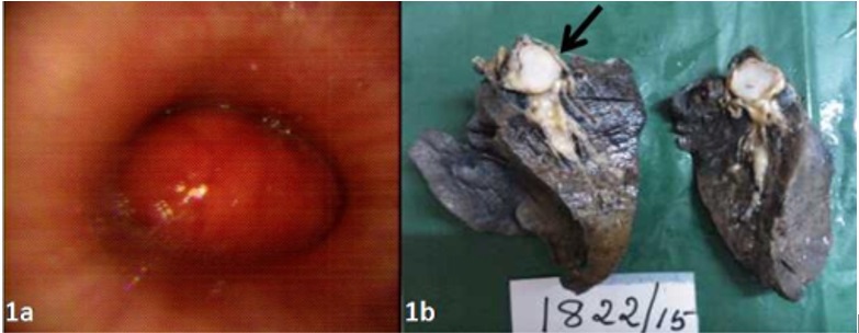
A 37 year old man was referred to our hospital with a 2 month history of breathlessness, cough and right sided pleural effusion. He was found to have an obstructive lesion in the right intermediate bronchus on chest x-ray and bronchoscopy (Fig. 1a). There was no submandibular or parotid gland enlargement. The laboratory studies, including the peripheral blood smear, biochemical examination, and abdominal ultrasound showed no abnormalities. A right lower bilobectomy (middle and lower) was done with a clinical diagnosis of Carcinoid – right bronchus.
Macroscopic examination showed a 2.8cm sized well circumscribed bluish white glistening homogenous unencapsulated mass in the right middle lobe bronchus (Fig. 1b).
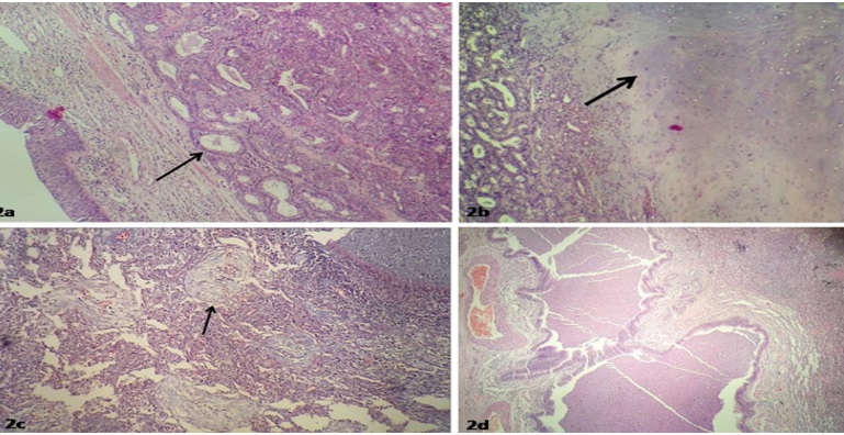
Microscopy showed a well circumscribed biphasic tumor made up of epithelial and mesenchymal components. Epithelial component was seen in the form of ductal lumina lined by cuboidal epithelium, surrounded by myoepithelial cells in sheets and nests (Fig. 2a) merging with large chondroid and myxoid areas (Fig. 2b).
Surrounding lung parenchyma showed distal air spaces filled with fibroblastic plugs (Masson bodies) (Fig. 2c), – a feature of Cryptogenic organising pneumonia, confirmed by Masson trichrome staining.
Thickened and dilated bronchioles with intraluminal inflammatory exudate – feature of post obstructive bronchiectasis was also seen (Fig. 2d).
Lower lobe showed obstructive emphysema with dilated alveoli showing thinning and destruction of alveolar septae. There was positivity of the epithelial ductal elements for the antibodies to cytokeratin and vimentin (Fig. 3a). The myoepithelial component and the mesenchymal component of the tumor showed positivity for vimentin (Fig. 3b).
On followup, recovery was uneventful and patient is alive and well after 8 months.
PPPA usually originate from the epithelium of the submucosal bronchial gland. They, therefore present as endoluminal lesions and rarely occur in peripheral or subpleural locations.[4] There is no gender or age predominance: the ages ranging between 8 and 74 years.[5]
PPPA are biphasic like their salivary gland counterpart, but do not feature either a prominent glandular component or chondroid stroma like the salivary gland counterpart. Rather, tumors exhibit features of the so called “cellular mixed tumor” manifesting sheets, trabeculae or islands of epithelial and/or myoepithelial cells and a myxoid matrix. The present case featured predominance of chondroid component an unusual finding in PPPA. Mitotic activity, pleomorphism and necrosis are unusual.[3] PPPA differ from pulmonary metastases of primary salivary gland tumors because they usually lack ducts or have very small branching ductules.[6]Positive cytokeratin and vimentin immunochemical staining of the pleomorphic adenoma epithelial elements supports the diagnosis as primary tumors. In contrast, cytokeratin staining is more prominent in the salivary gland counterpart.6 Differential diagnosis includes chondroid hamartoma, pulmonary blastoma and carcinosarcoma. Chondroid hamartomas usually show mature cartilage that predominate the lesion while the latter tumors feature obvious malignant stroma and epithelium.[6]
Approximately one third of cases are found incidentally.[7] The majority of patients do present with symptoms like cough, sputum production, dyspnea wheeze and stridor.[8] In the present case due to the presence of intraluminal obstructive lesion of the bronchus, the surrounding lung showed cryptogenic organising pneumonia with post obstructive bronchiectasis and the lower lobe showed obstructive emphysema.
In conclusion PPPA is an extremely rare tumor and metastasis from the salivary gland should be ruled out by thorough clinical history and examination. This case of endoluminal pulmonary pleomorphic adenoma is interesting because of coexisting cryptogenic organising pneumonia and post obstructive bronchiectasis. Immunohistochemical staining supports the diagnosis of primary lung neoplasm. The treatment for this tumor is still surgical excision.[6]
 |
Click here to view |
Fig. 1: (a) Flexible bronchoscopy intraluminal mass seen at the right intermediate bronchus. (b) Cut surface shows a bluish white glistening homogenous mass in the right middle lobe bronchus.
 |
Click here to view |
Fig. 1: (a) Flexible bronchoscopy intraluminal mass seen at the right intermediate bronchus. (b) Cut surface shows a bluish white glistening homogenous mass in the right middle lobe bronchus.
 |
Click here to view |
Fig. 3: (a) Cytokeratin positivity in the epithelial component. (b) Positive vimentin stain seen in the mesenchymal component.
How to cite : Lal L, Philipose T R, Samartha V, Primary pulmonary pleomorphic adenoma with cryptogenic organising pneumonia – A rare entity. IP J Diagn Pathol Oncol 2019;4(1):78-80
This is an Open Access (OA) journal, and articles are distributed under the terms of the Creative Commons Attribution-NonCommercial-ShareAlike 4.0 License, which allows others to remix, tweak, and build upon the work non-commercially, as long as appropriate credit is given and the new creations are licensed under the identical terms.
Viewed: 1575
PDF Downloaded: 559