- Visibility 66 Views
- Downloads 12 Downloads
- DOI 10.18231/2581-3706.2019.0014
-
CrossMark
- Citation
Histomorphological analysis of uterine and cervical lesions in hysterectomy specimens at a tertiary care hospital
- Author Details:
-
Sujatha R
-
Manjunatha Y. A
-
Jaishree T *
Abstract
Introduction: The Uterus is a major female hormone responsive reproductive organ which is subjected to vast range physiological changes, benign and malignant disorders.
Aims and Objectives: The study was undertaken to see different patterns of histomorphological lesions in uterus and cervix in hysterectomy specimens and to study its correlation with respect to age and clinical presentation.
Materials and Methods: This study is a prospective analysis of 155 hysterectomy specimens received in Department of Pathology in DR B R Ambedkar medical college and hospital in Bangalore during a 18 months period from June 2017 to December 2018.
Results: In the present study commonest type of hysterectomy was Total abdominal hysterectomy and the commonest clinical indication being Menorrhagia in 63(40.64%) cases. The peak age of incidence of hysterectomy was noted in the 5th decade (38.91%). The commonest lesion encountered was Chronic non specific cervicitis in 86(55.40%) cases and leiomyoma in 57(36.77%) cases, 6 cases (3.87%) of carcinoma endometrium, 9 cases (5.80%) of carcinoma cervix and 11 case (7.00%) of cervical dysplasia were encountered. The rare and interesting lesions encountered in the present study were 2(1.28%) case of blue nevus Cervix and 2 case (1.28%) of Benign Cellular Leiomyoma and 1 case of (0.64%) Lymphoepithelioma like SCC cervix
Conclusion: Hysterectomy still remains one of the most commonly done procedure in the gynecology department. Histomorphological analysis of lesions of uterus and cervix is mandatory for establishing final diagnosis.
Keywords: Histomorphology, Uterus, Cervix, Hysterectomy, Menorrhagia, Leiomyoma and carcinoma cervix, SCC.
Introduction
The female reproductive tract is a complicated but fascinating system comprising of the external and the internal genitalia with vagina, uterus, fallopian tubes and ovaries. Uterus, additionally called the womb and the cervix are the vital female reproductive organs, subjected to numerous non neoplastic and neoplastic diseases. The uterus consists of the endometrium and the myometrium, which are continuously stimulated by hormones, denuded monthly of its endometrial mucosa and inhabited periodically by fetuses.[1] Together with the lesions that affect the cervix, the lesions of the corpus of the uterus and the endometrium account for most patient visits to gynecologists.[2]
Hysterectomy is the surgical removal of the uterus and is performed by a gynecologist. Historically Charles Clay performed the first subtotal hysterectomy in Manchester England in 1843 and the first Total abdominal hysterectomy was done in 1929.[3] Hysterectomy still remains one of the most prevalent gynecological procedure performed worldwide in spite of other treatment modalities for many gynecological disorders.
This study is entitled to study and understand the various gross and histomorphological findings in the uterus and cervix of hysterectomy specimens and their distribution with respect to age of the patient and the mode of presentation.
Materials and Methods
This prospective type of study was carried out in the Department of Pathology, Dr. B.R. Ambedkar medical college and hospital, Banglore over a period of 18 months between ‘Jun 2017 to Dec 2018’.There were 155 hysterectomy specimens received from gynecology department included in this study. Hysterectomy specimens with indications for pathologies related to fallopian tubes and ovaries were omitted from the study. Clinical details of the patients were obtained from the requisition forms received along with the hyssterectomy specimens and were entered in the proforma.
All the received hysterectomy specimens were immediately fixed with 10% formalin saline in the ratio of 1:10. After 24 hours of fixation, the specimens were thoroughly examined grossly and then multiple representative bits were taken, processed and embedded in paraffin. Three to five micron thickness sections were taken; slides were made and stained with routine H & E stain. The histomorphological findings of uterus and cervix were noted and then these findings were correlated with age and clinical diagnosis.
Results
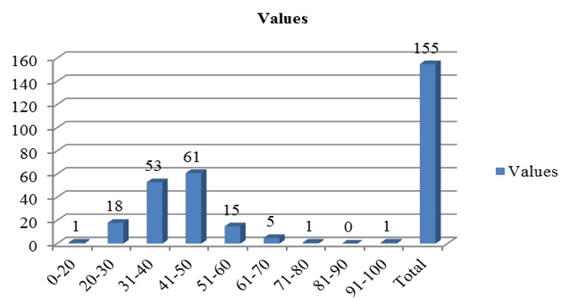
A total of 155 hysterectomy specimens were analyzed in this study. The incidence of hysterectomies in relation to the age was studied and depicted in the Fig. 1. Age ranged from 18 years to 97 years with mean age of 41.78. In our present study, peak age of hysterectomy done was found in 5th decade i.e 41 – 50 yrs with 61 of cases. (Fig. 1) The commonest type of the hysterectomy encountered was total abdominal hysterectomy in 66(42.75%) cases. (Table 1)
 |
Click here to view |
Fig. 1: Age distribution of all the hysterectomy specimens.
 |
Click here to view |
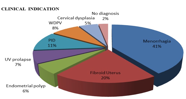
Fig. 2: Clinical indications of all the hysterectomies
The various clinical indications for hysterectomies were analyzed and depicted in Fig. 2. Most of the patients clinically presented with Menorrhagia which was seen in 66 cases (40.64%), and was followed by Fibroid uterus in 31 cases (20%).
Table 1: Distribution of case as per the type of hysterectomy
|
Type of Hysterectomy |
Number of cases |
|
Partial Hysterectomy |
27(17.4%) |
|
Vaginal Hysterectomy |
19(12.2%) |
|
Total Abdominal Hysterectomy |
66(42.5%) |
|
Total Hysterectomy with Bilateral Salphingo-oophrectomy |
43(27.7%) |
|
Total |
155 |
The histomorphological findings were analyzed.
In the endometrium, various findings encountered were, proliferative phase in 83 cases (53.54%) was the commonest finding followed by simple hyperplasia in 31 cases (20.0%). Atrophic pattern in 9 cases (5.80%),
endometritis in 4 cases (2.58%), Complex hyperplasia without atypia in 3 cases (1.93%), endometrial polyp in 7 cases (4.51%) and endometrial carcinoma in 6 cases (3.87%). Pill endometrium (Iatrogenic change) was seen in 2 cases (1.29%). (Table 2).
Table 2: Endometrial change seen in all the 155 specimens.
|
S.no |
Endometrial Change |
Number of cases |
|
1. |
Proliferative Endometrium |
83(53.54%) |
|
2. |
Secretory Endometrium |
10(6.45%) |
|
3. |
Atrophic Endometrium |
09(5.80%) |
|
4. |
Simple Hyperplasia |
31(20.0%) |
|
5. |
Complex hyperplasia |
03(1.93%) |
|
6. |
Endometrial Polyp |
07(4.51%) |
|
7. |
Endometritis |
04(2.58%) |
|
8. |
Pill endometrium |
02(1.29%) |
|
9. |
Carcinoma Endometrium |
06(3.87%) |
|
|
Total |
155 |
In the myometrium, most common finding encountered was Leiomyoma in 66 cases (42.58%) followed by adenomyosis in 35 cases (22.58%). Myometrial hyperplasia was encountered in 2 cases (1.29%). In case of leiomyomas, 2 cases (1.29%) were benign cellular leiomyoma and 7 cases (4.51%) showed degenerative changes. (Table 3)
Table 3: Myometrial lesions seen in all the 155 specimens.
|
S.no |
Myometrial changes |
Number of cases |
|
1. |
Unremarkable |
21(13.54%) |
|
2. |
Leiomyoma |
66(42.58%) |
|
3. |
Adenomyosis |
35(22.58%) |
|
4. |
Leiomyoma with adenomyosis |
26(16.77%) |
|
5. |
Monckebergs’ Calcific sclerosis |
05(3.22%) |
|
6 |
Myometrial Hyperplasia |
02(1.29%) |
|
|
Total |
155 |
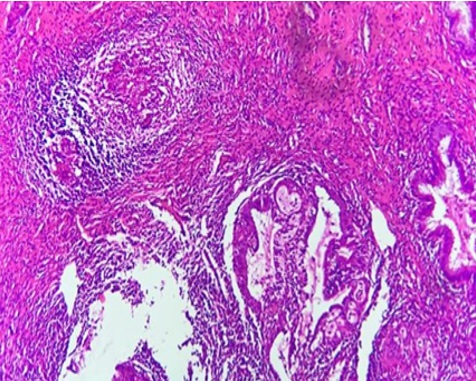
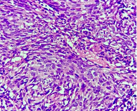
The histomorphological study of cervix showed Chronic non specific cervicitis as most common finding seen in 86 cases (55.40%). Granulomatous cervicitis seen in 2 cases (1.20%), polypoidal endocervicitis in 16 cases (10.30%) and microglandular hyperplasia in 03 cases (1.90%). 4 cases (2.50%) of endocervical polyp and 2(1.20%) cases of blue nevus were also encountered. In our study 11 cases (7.00%) of cervical dysplasias and 9 cases (5.80%) of carcinoma cervix were noted. In case of Cervical carcinoma 6 cases were of Squamous cell carcinoma cervix, 1 case of Adenocarcinoma cervix, 1 case of Adenosquamous carcinoma cervix and 1 case was Lymphoepitheilioma like SCC.
Table 4: Histomorphological lesions of cervix.
|
S.no |
Histomorphological Findings |
Number of cases |
|
1. |
Chronic non specific cervicitis |
86(55.40%) |
|
2. |
Polypoidal endocervicitis |
16(10.30%) |
|
3. |
Granulomatous Cervicitis |
02(1.20%) |
|
4. |
Microglandular hyperplasia |
03(1.90%) |
|
5. |
Cervical Polyp |
04(2.50%) |
|
6 |
Cervical Fibroid |
02(1.20%) |
|
7. |
Blue nevus – Cervix |
02(1.20%) |
|
8. |
Chronic non specific cervicitis with Squamous metaplasia |
20(12.90%) |
|
9. |
Cervical Dysplasia |
11(7.00%) |
|
10 |
Carcinoma Cervix |
09(5.80%) |
|
|
Total |
155 |
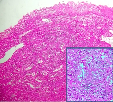
Rarest and interesting lesions encountered were 1 case of lymphoepithelioma like SCC, 2 cases of Benign Cellular leiomyoma and 2 cases of Blue nevus.
In 21 hysterectomy specimens no pathological findings were encountered.
Discussion
Hysterectomy is the most commonly performed major gynecological surgery throughout the world. It is a successful operation in terms of symptom relief and patient satisfaction and provides definitive cure to many diseases involving uterus as well as adnexa.[4]
This study was undertaken to analyze and understand the histomorphological spectrum of uterine and cervical lesions in hysterectomy specimens and to determine its correlation with respect to age and clinical diagnosis of the patient.
In our study commonest age group for hysterectomy specimens was 5th decade, which is in concordance with studies by Patil HA et al,[5] Ajmera Sachin K et al[6] and Yogesh Neena et al.[7]
In our study, the incidence of abdominal hysterectomy was higher than the vaginal as in the studies done by Ajmera Sachin K et al,[6] Sunita Malik et al[8] and Ankur gupta et al.[9]
The present study revealed Menorrhagia as most common indication which was found to be in concordance with studies done by Ticku et al[10] and Karthikeyan et al.[11]
Endometrial polyp in this present study was 7 cases (4.51%) which was seen to be in concordance with study conducted by Yogesh Neena et al which was 3.47%.[7]
In this present study endometrial hyperplasia accounts to 34 cases (21.9%). This finding was found comparable with the studies conducted by Ojeda VJ [12]and Ranabhat et al,[13] which showed 22.3% and 16% cases of endometrial hyperplasia respectively.
In our study endometrial carcinoma was encountered in six cases (3.80%), and mainly found in post menopausal age group presenting with post menopausal bleeding. The study done by Ankur gupta et al[9] has similar findings.
Among the myometrial lesions, leiomyoma accounts for majority comprising of 66 cases (42.58%), which is in comparison with studies done by Ajmera Sachin K et al[6]in 170 cases (48.6%) and Stewart and Arti.[14] Majority of the studies done on the histopathological analysis of hysterectomy specimens so far reveals that uterine fibroids are the most common pathology to be encountered in the uterus.
Adenomyosis (22.58%) was second most common pathology in our present study which was in accordance with study conducted by Sunita malik et al[8] showing 21%. Adenomyosis is very rare and difficult to diagnose pre operatively as it has no specific clinical symptom of its own. It is usually diagnosed only after hysterectomy by histomorphological examination of the specimen. Around 26 cases (16.77%) in this study revealed the presence of both leiomyoma and adenomyosis.
Non neoplastic diseases of uterine cervix are predominantly inflammatory and are more common. A vast majority of cases with inflammatory pathology had no specific cause and chronic non specific cervicitis being the most common lesion encountered accounting to 55.40% which is in comparable with other studies done by Nwachokor & Forae[15] and Garewal et al.[16]
 |
Click here to view |
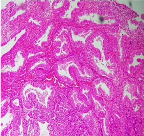
Fig. 3: Complex hyperplasia of Endometrium: Micrograph shows crowded branching and papillary glands seperated by scant stroma. (H & E at 40X).
 |
Click here to view |
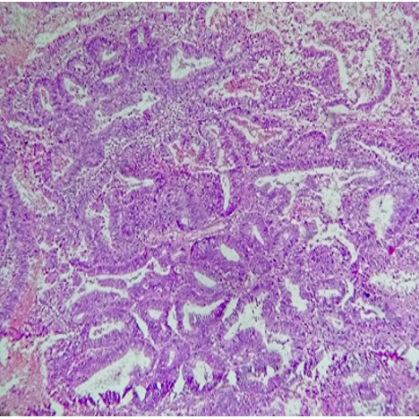
Fig. 4: Endometrial Adenocarcinoma: Micrograph shows atypical crowded glands with invasion into the stroma. (H & E at 40X).
Among the myometrial lesions, leiomyoma accounts for majority comprising of 66 cases (42.58%), which is in comparison with studies done by Ajmera Sachin K et al[6]in 170 cases (48.6%) and Stewart and Arti.[14] Majority of the studies done on the histopathological analysis of hysterectomy specimens so far reveals that uterine fibroids are the most common pathology to be encountered in the uterus.
 |
Click here to view |
Fig. 5: Granulomatous Cervicitis: Micrograph shows well formed granulomas adjacent to endocervical glands.(H & E at 10x).
 |
Click here to view |
Fig. 6: Poorly differentiated SCC: Mirograph shows squamous cells with high N: C ratio, marked atypia and minimal keratinization. (H & E at 40X).
Adenomyosis (22.58%) was second most common pathology in our present study which was in accordance Intraepithelial neoplasia is a very critical in the the natural history of cervical carcinoma. The importance of detecting and treating patients at this stage is that the progression of intraepithelial neoplasia into carcinoma can be prevented.In this present study cervical dysplasia accounts for 9 cases (5.80%) which is in comparison with Vijaya Gattu et al[17] and Cervical cancer accounts for 8 cases (5.16%) which is also in concordance with Vijaya Gattu et al.[17]
Adenomyosis (22.58%) was second most common pathology in our present study which was in accordance with study conducted by Sunita malik et al[8] showing 21%. Adenomyosis is very rare and difficult to diagnose pre operatively as it has no specific clinical symptom of its own. It is usually diagnosed only after hysterectomy by histomorphological examination of the specimen. Around 26 cases (16.77%) in this study revealed the presence of both leiomyoma and adenomyosis.
Non neoplastic diseases of uterine cervix are predominantly inflammatory and are more common. A vast majority of cases with inflammatory pathology had no specific cause and chronic non specific cervicitis being the most common lesion encountered accounting to 55.40% which is in comparable with other studies done by Nwachokor & Forae[15] and Garewal et al.[16]
 |
Click here to view |
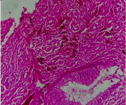
Fig. 7: Blue nevus cervix: Micrograph shows wavy dendritic cells arranged in clusters next to endocervical gland. (H & E at 10X).
Uterine cervix is a very unusual site for pigmented melanotic lesions.Blue nevi are infrequent incidental findings encountered in the cervix of hysterectomy specimens from middle aged females. In our study 2 cases (1.20%) of blue nevus cervix was encountered as a incidental finding in the hysterectomy specimen
 |
Click here to view |
Fig. 8: Lymphoepithelioma like SCC: Micrograph shows syncytium of poorly differentiated tumour cells with prominent lymphoplasmacytic infiltrate.(H& E at 10X).
Conclusion
Hysterectomy still remains one of the most common treatment modality worldwide, thereby forming substantial work load in the histopathology department. This present study provides a limelight on the various histomorphological findings of uterus and cervix encountered in hysterectomy specimens at our institution. The histomorphological analysis was found to correlate well with the clinical diagnosis of the patients and quite a few lesions were also encountered completely as pure incidental findings. Hence, it becomes mandatory that every hysterectomy specimen, even if it is grossly appearing to be normal, it should be subjected to exhaustive histomorphological examination so as to ascertain better post operative management and follow up.
Conflict of Interest: None
References
- ^ Ellenson LH, Pirog EC. The female genital tract.In: Kumar V, Abbas AK, Fausto N, Aster JC. Robbbins and Cotran Pathologic basis of disease.8th ed. Philabelthia:Elsevier 2010:1005-1063.
- ^ Qamar-Ur-Nisa, Habibullah, Shaikh TA, Hemlata, Memon F, Memon Z. Hystrectomies; An audit at a tertiary care hospital. Prof Med J 2011;18(1):46–50.
- ^ John, A., Rock, M. D., Jhon, D., & Thompson, M.D. (2003). Telind’s Operative Gynaecology. 1st edition Lipincott –Ravenplace.
- ^ Jaleel R, Khan A, Soomro N. Clinicopathological study of abdominal hysterectomies. Pak J Med Sci 2009;25(4):630-34
- ^ Harshal A. Patil, Archana Patil and Suresh V. Mahajan. Histopathological Findings in Uterus and Cervix of Hysterectomy Specimens. MVP J Med Sci 2015;2(1):26–29.
- a, b, c, d Ajmera SK, Mettler L, Jonat W. Operative spectrum of hysterectomy in a German university hospital. J Obstet Gynecol India 2006;56(1):59–63.
- a, b Neena Y, Honey B, Nema SK, Kural MR. clinicopathological correlation of hysterectomy specimens for abnormal uterine bleeding in rural area. JEMDS 2013;39(1):7506-7512.
- a, b, c Sunita Malik, Jai Bhagwan Sharma, Nirmal Gulati, and K.Jain. A Clinico- Pathological Study of Adenomyosis. J Obstet Gynecol India 1992;42:234-238.
- a, b Ankur Gupta, Sangita Sehgal, Ajay yadav, Vinod Kumar. Histopathological Spectrum of Uterus and Cervix in Hysterectomy Specimens. Int J Med Res Prof 2016;2(3):136-139.
- ^ Dr. Prem Singh, Dr. Archana Ticku and Dr. Gitanjali Jamwal. Histopathological spectrum of hysterectomy specimens in tertiary care hospital: a prospective study. ejbps, 2017;4(8):858-866.
- ^ Karthikeyan, T. M., Veena, N. N., & Ajeeth Kumar, C. R. (2015). Clinicopathological study of hysterectomy among rural patients in a tertiary care center. IOSR J Dent Med sci 14(1):25-27.
- ^ Ojeda VJ. The pathology of hysterectomy specimens. Z Med J 1979;89(631):169-1671;1(1):2
- ^ Ranabhat SK, Shrestha R, Tiwari M, Sinha DP, Subedee LR. A retrospective histopathological study of hysterectomy with or without salpingo-ophorectomy specimens. JCMC 2010;1(1):26–29.
- a, b Stewart FC and Arti P. The Frequency of Uterine Leiomyomas. Am J Clin Pathol 1990;94:435-438.
- a, b Nwachokor FN and Forae GD.Morphological spectrum of non-neoplastic lesions of the uterine cervix in Warri, South-South, Nigeria. Niger J Clin Pract 2013 Oct-Dec;16(4):429-432.
- a, b Garewal j, khatri L S, Saxena V,.Clinicopathological Evaluation of NonNeoplastic and Neoplastic Lesions of Uterine Cervix. Imperial J Interdiscip Res (IJIR) 2016;2(4):426-430.
- a, b Vijaya Gattu. Histomorphological Study of Uterus and Cervix and Correlation with Clinical Diagnosis. Sch J App Med Sci 2016;4(12B):4285-4290.
