Author Details :
Volume : 4, Issue : 1, Year : 2019
Article Page : 39-44
https://doi.org/10.18231/2581-3706.2019.0007
Abstract
Introduction: Like any other human organ, kidney also may be involved by many inflammatory, benign or malignant diseases which require removal of kidney. Simple nephrectomy is done to remove the irreversibly damaged, non-functioning kidneys involved by different benign pathologic condition while radical nephrectomy is indicated in malignant lesion.
Materials and Methods: The materials for the study were collected from the patients being admitted in a tertiary care hospital situated in Ahmedabad affiliated with Gujarat University. Data were collected in pretested proforma.
Results: Total 74 cases of nephrectomy specimen were studied. The data according to age incidence, sex incidence, location, nature of the lesion and clinical symptoms was prepared and analyzed. 55 (74.33%) cases were of benign lesions while 19 (25.67%) cases were of malignant lesions. We found 6 cases of congenital kidney diseases, among which maximum cases were of multicystic renal dysplasia (50%), followed by bilateral renal dysplasia, horse shoe kidney and duplex kidney (16.66% each).
Conclusion: Most common benign lesions were Chronic inflammatory causes, renal calculus related hypofunctioning kidneys, trauma and congenital diseases of kidney. Angiomyolipoma and Cystic nephroma were most common benign neoplasm while renal cell carcinoma, Wilm’s tumor and urothelial carcinoma were most common malignant neoplasm.
Keywords: Nephrectomy specimen, Benign, Malignant, Congenital lesion.
Like any other human organ, kidney also may be involved by many inflammatory, benign or malignant diseases which require removal of kidney. Simple nephrectomy is done to remove the permanently damaged, not working kidneys involved by different benign pathological conditions like extensive renal calculous, obstruction in ureter, or at pelviureteric junction (PUJ), On the other hand, radical nephrectomy is indicated in malignant lesion. A wide variety of both benign and malignant tumors are found in kidney.[1]For accurate diagnosis histopathology evaluation of renal tumor is necessary. Moreover, currently the ideal standard in the treatment of all tumors of kidney is radical or partial nephrectomy.[2] Histopathologic examination of tumor in nephrectomy specimens is essential to establish histologic type and to record accepted histopathological prognostic markers like i.e. tumor size, histological subtype, nuclear grade, and stage in cases of malignant lesion.[2]
In the recent time, there has been a growing interest on kidney saving surgery or partial nephrectomy to treat localized malignant lesion by laparoscopic approach.[3]In the developed countries, laparoscopic procedures have outperformed open nephrectomy procedures.[4]But in our country, in many centers, especially those in rural or semi-urban setup, we have been performed open nephrectomy procedures.
Material and Methods
Source and Method of collection of data
This was a three and a half year study conducted in histopathology section of Department of Pathology, NHL Municipal medical college Ahmedabad from June 2009 to October 2012. All patients who visited the Surgery/Urology outpatient department and presenting with haematuria, dysuria etc., were included in the study. Simple or radical nephrectomies were performed as and when indicated. During the chosen study period total of 74 patients went under nephrectomy.
Nephrectomy specimens were received and fixed in 10% buffered formalin. Grossing of nephrectomy specimens was done according to the standard protocol.[5] Representative tissue were taken and processed for paraffin embedding, haematoxylin and eosin (H and E) stained and examined by the pathologists. Special stains (modified Z.N. stain and PAS stain) were done whenever required. Pathological staging was performed with the 2002 TNM classification[6] and histological subtyping was performed by the 2004 WHO classification.[7]Each tumor was graded according to the Fuhrman nuclear grading system.[8]
Sample size
All the nephrectomies which were done in our hospital from June 2009 to October 2012 were part of our study.
A total of 74 patients underwent nephrectomy during the three and half year period. Age of the patients were ranging from 1 day to 80 years (mean age 38.34±22.28years)
Table 1: Distribution of neoplastic and non neoplastic lesions by age and sex
|
Age (yrs) |
Benign and Malignant Lesion |
Non neoplastic lesions |
Total |
Percentage (%) |
||
|
Male |
Female |
Male |
Female |
|||
|
0-1 |
1 |
0 |
3 |
2 |
6 |
8.11% |
|
01-10 |
2 |
1 |
3 |
2 |
8 |
10.81% |
|
11-20 |
0 |
0 |
2 |
3 |
5 |
6.75% |
|
21-30 |
1 |
0 |
5 |
0 |
6 |
8.11% |
|
31-40 |
0 |
1 |
6 |
3 |
10 |
13.51% |
|
41-50 |
1 |
2 |
9 |
3 |
15 |
20.27% |
|
51-60 |
3 |
7 |
5 |
2 |
17 |
22.97% |
|
61-70 |
2 |
0 |
1 |
1 |
4 |
5.40% |
|
>70 |
1 |
1 |
1 |
0 |
3 |
4.05% |
|
Total |
11 |
12 |
35 |
16 |
74 |
100% |
Maximum nephrectomies were done in age group51-60 years (17 cases; 22.97%). Also nephrectomies for malignant lesions were highest in this age group (7 cases; 36.84%).
Table 2: Sex predilection in neoplastic and non neoplastic lesions
|
No. of nephrectomy cases (%) |
Male |
Female |
|
Total no. of cases =74(100%) |
46 |
28 |
|
Neoplastic conditions = 23(31.1%) Benign Neoplasm(4) Malignant Neoplasm (19) |
11 0 11 |
12 4 8 |
|
Non neoplastic conditions =51(68.9%) |
35 |
16 |
Out of 74 nephrectomy specimens, Male Female ratio was 1.63:1
35 (47.29%) kidneys removed were from the left side while 37 (50%) kidneys were of right side from total cases of 74 [Table 3]. One case of bilateral kidneys were removed for renal dysplasia. In another case horse shoe kidney was the indication for nephrectomy.
Table 3: Histopathological diagnosis
|
Diagnosis |
No. of patients |
Percentage (%) |
|
Benign conditions Congenital Inflammatory Traumatic Benign neoplasms |
55 6 39 6 4 |
74.33% 7.89% 52.70% 7.89% 5.40% |
|
Malignant neoplasms |
19 |
25.67% |
|
Total |
74 |
100% |
Out of 74 cases, 55 (74.33%) cases were of benign conditions while 19 (25.67%) were of malignant conditions of the kidney [Table 3].
Within the non neoplastic lesions, 39 cases were of Chronic inflammatory lesion followed by trauma (6 cases) and congenital diseases of kidney (6 cases).
Within malignant lesion Renal cell carcinoma (14 cases;73.68%) was the most common etiology.
Among the 4 cases of benign neoplasms 3 (75%) cases were of Angiomyolipoma and 1(25%) case was of Cystic nephroma.
We found six cases of congenital kidney diseases, among which maximum cases were of multicystic renal dysplasia(3 cases; 50%), followed by bilateral renal dysplasia, horse shoe kidney and duplex kidney one case each(16.66%).
Table 4: Histological Types of Renal Cell Carcinoma (RCC)
|
RCC Types |
Observations |
Percentage (%) |
|
Clear cell RCC |
8 |
57.14% |
|
Papillary RCC |
2 |
14.29% |
|
Chromophobe RCC |
3 |
21.42% |
|
Multilocular cystic RCC |
1 |
7.14% |
|
Total |
14 |
100% |
Conventional (clear cell) Renal cell carcinoma was the leading variant of Renal cell carcinoma in our study (8 cases; 57.14%). We found 3 cases(14.29%) of Chromophobe type; and 2 cases(14.29%) of Papillary Renal cell carcinoma and 1 case of multilocular cystic Renal cell carcinoma.
Table 5: Histopathologic Characteristics of 8 cases of Clear Cell Renal cell carcinoma
|
Histopathologic Characteristics |
No. of Clear cell RCC cases |
Percentage (%) |
|
Capsular Invasion |
3 |
37.5% |
|
Renal Vein Invasion |
2 |
25% |
|
Renal Sinus Invasion |
2 |
25% |
|
Perinephric fat invasion |
2 |
25% |
|
Fascia of Gerota invasion |
1 |
12.25% |
|
Staging pT1 pT2 pT3 pT4 |
3 2 2 1 |
37.5% 25% 25% 12.25% |
|
Fuhrman’s Nuclear grading Grade I Grade II Grade III Grade IV |
1 5 1 1 |
12.25% 62.5% 12.25% 12.25% |
The main pathologic prognostic parameters which are important in Clear Cell RCC, are mention in Table-5.The maximum diameter of primary tumor was 16 cm, as seen in 1 case (12.25%) and the least 1.5 cm, seen in 1case (12.25%). Capsular invasion was seen in 3 cases (37.5%). Renal vein invasion was seen in 2(25%) cases, while renal sinus invasion present microscopically in 2(25%) cases. Involvement of perinephric fat was observed in 2 cases
(25%) While Fascia of Gerota was involved in one case (12.25%) only. Lymph nodes were received in 3(37.5%) cases. Lymph node metastases were not present in any case.
Among 8 cases of Clear Cell RCC, one case (12.25%) is of grade I, 5 cases (62.5%) are of grade II, one case (12.25%) is of grade III and one case (12.25%)is of grade IV.
 |
Click here to view |
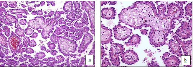
Fig 1: Papillary Renal cell carcinoma: H and E stain (10x), Fig 2: Papillary renal cell carcinoma: H and E stain (40x)
 |
Click here to view |
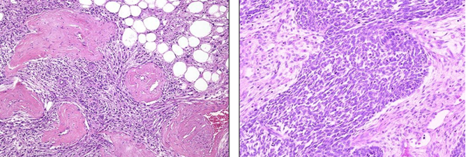
Fig. 3: Angiomyolipoma of Kidney_showing angio adipose tissue, smooth muscle, and thick-walled blood vessels H & tain, Fig. 4: Wilm’s tumor: H & E stain (40x)
Discussion
There is definitely a geographic disparity regarding the indications of nephrectomy. In our study, there is a much higher rate of nephrectomy performed for non neoplastic conditions of the kidney compared to developed country. In our present series of 74 nephrectomies performed during the study period of three and half years, 74.33% cases were performed for benign conditions whereas 25.67% cases were performed for malignant diseases of the kidney. which is comparable with the series reported from developing countries.
Table 6: Incidence of benign V/S malignant lesions
|
Various Studies |
Benign lesions (%) |
Malignant lesions (%) |
|
Nigeria(Eke N.Echem)[9] |
32.70 |
67.30 |
|
Norway(Beisland C.)[10] |
32.00 |
68.00 |
|
Korea(Badmus TA)[11] |
42.88 |
57.12 |
|
Philips et al[12] |
24.70 |
75.30 |
|
Sudan(Ghalayini et al)[13] |
70.00 |
30.00 |
|
Saudi Arabia(Malik et al)[14] |
77.60 |
22.40 |
|
Pakistan(Rafique M. et al)[15] |
76.60 |
23.40 |
|
India(Darjeeling,Datta et al)[16] |
60.20 |
39.80 |
|
Present study |
74.33 |
25.67 |
Out of total 74 nephrectomies 46 (62.1%) patients were male and 28 (37.9%) were females (Male: Female ratio 1.63:1). Male:female ratio was comparable to Eke N.et al,[9]Mahesh Kumar et al,[17] Fauzia Latif et al,[18] Datta et al.[16]In Rafique M. et al[15] series male female ratio was 1:1.06. [Table 6] Age of the patients ranged from 1day to 80 years (Mean age 38.34±22.28 years).
Pyelonephritis was the leading pathological entity in our nephrectomies (52.7%), which is compatible with the report by Kubba, et al,[19] and Malik et al[14]but different from that of Schiff and Glazier[20]. In these latter studies, RCC and TCC combined were the leading pathological findings.
Six(8%) of the removed kidneys in our series contained stones, which is comparable to the 6% in a large adult series of nephrectomies by Schiff and Glazier[20] and 11% in a pediatric series by Adamson et al and different from the 37% in Malik et al series[14]
Renal tuberculosis is an infection worth of mentioning in India as three cases (5.45%) of tuberculosis were found in our study among the nephrectomies carried out for benign conditions as compared to 7.62% in the report of Rafique M. et al[15] and 16.36% in report of Datta et al[16] In Kubba, et al[19]and Malik et al[14]there were no tuberculosis cases. Beisland et al[10] found that five (2.4%) tuberculous kidneys were removed out of 209 nephrectomies carried out for benign conditions during 20 years at two Norwegian hospitals. Another report from Ghalayini et al[13] showed that tuberculosis accounted for nine (3%) nephrectomies performed for benign conditions, whereas patients with renal tuberculosis are uncommon in developed countries.
Renal tumors in adults are increasing in incidence throughout the world, partly as a result of widespread use of cross sectional imaging modalities and ultrasonography. Both benign and malignant tumors occur in the kidney. Because of the relative rarity of benign renal tumors, it is a common practice for urologists to consider any renal mass that enhances with intravenous contrast on computed tomography (CT) scan as a malignancy. If it is localized,
they tend to treat such masses radically unless there is
definite evidence of a benign pathology. Most common malignant tumor in adults is renal cell carcinoma (RCC) and Wilm’s tumor in childhood. Rare are urothelial tumors of calyces and pelvis.
Of 23 renal tumors in this series four (17.3%) were benign as compared to a recent report on Saudi patients by Talik RF et al[21]where 14% of renal tumors were benign and Mahesh Kumar et al39 where 16.6% cases were benign.This is in marked contrast with 5% of Malik et al,[14] 0% of Rafique M. et al,[15] 8% of Datta et al[16] and 6% of Fauzia Latif et al[18]
RCC is the most common primary malignant tumor of the kidney (85%) worldwide and constitutes 2-3% of all visceral malignancies in adults. The classification of renal cell neoplasms has been extensively studied in the last two decades and is based on a combination of histological, genetic and immunohistochemical features.
The incidence of renal cell carcinoma according to Datta et al[16]is about 69.6%, Fauzia Latif et al[18] is 87.2%, Rafique M. et al[15] is 100%, Eble et al[22]is about 90%, McLaughlin JK et al[23] is about 85% and in our study it is 73.68%.
Fuhrman's nuclear grading system was applied in our study. Nuclear grade II (5 cases 62.5%) was the most common presentation, while both grade I and higher grades (III and IV) were rare in our study. The results were similar to Fauzia Latif et al.[18] Which noted 63.3% cases in grade II, while both grade 1 and higher grades (III and IV) were rare.
Conclusion
This was a three and a half year study of 74 nephrectomy specimens in histopathology Department NHL Municipal medical college Ahmedabad from June 2009 to October 2012.
How to cite : Shah N, Goyal S, Histopathological study of nephrectomy specimens in a tertiary care hospital. IP J Diagn Pathol Oncol 2019;4(1):39-44
This is an Open Access (OA) journal, and articles are distributed under the terms of the Creative Commons Attribution-NonCommercial-ShareAlike 4.0 License, which allows others to remix, tweak, and build upon the work non-commercially, as long as appropriate credit is given and the new creations are licensed under the identical terms.
Viewed: 3469
PDF Downloaded: 646